Korean J Phys Anthropol.
2017 Mar;30(1):15-20. 10.11637/kjpa.2017.30.1.15.
Morphometric Study of Cricoid Cartilage in Korean
- Affiliations
-
- 1Department of Anatomy, Chonbuk National University Medical School and Institute for Medical Sciences, Chonbuk National University, Cheongju, Korea. asch@jbnu.ac.kr
- KMID: 2374772
- DOI: http://doi.org/10.11637/kjpa.2017.30.1.15
Abstract
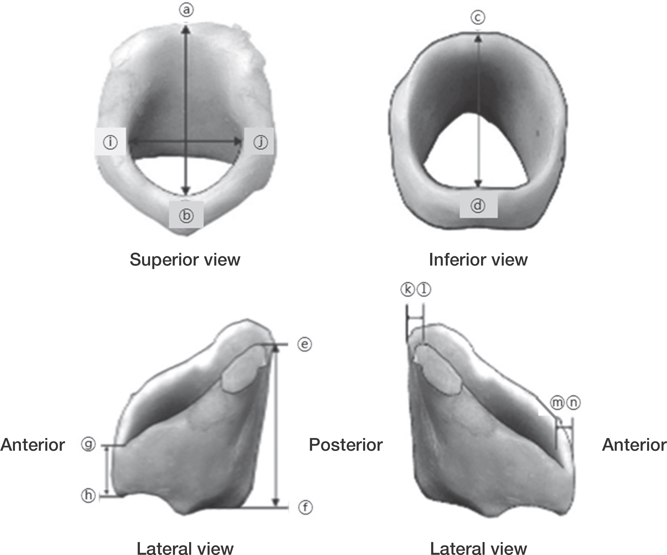
- This study is aimed to measure the morphology of Korean cricoid cartilages. A total of 48 - 33 males and 15 females - cadavers were used in this study. When it comes to their average age, males were 70 years old (50 to 91 years old), and females were 74 years old (47 to 92 years old). For this study, anteroposterior diameter and transverse diameter of superior side, anteroposterior diameter of inferior side, height of arch and lamina, anterior and posterior thickness of cricoid cartilages were measured. Anteroposterior diameters of superior and inferior cricoid cartilage were 28.5, 18.78 mm in male, and 23.85, 15.97 mm in female, respectively. And transverse diameters of superior side were 17.19 mm in male and 13.36 mm in female. Heights of arch and lamina were 7.10, 22.33 mm in male and 5.72, 20.10 mm in female, respectively. Thickness of anterior arch and posterior lamina were 2.57, 2.83 mm in male and 2.22, 2.42 mm in female, respectively. As a result, most Korean male measurements were significantly longer than female measurements except the anterior and posterior thickness of cricoid cartilages. Moreover the majority of measurements were shorter than Nigerians or Europeans. However, they were very similar to American Indians' measurements. In conclusion this study stated above can be a valuable foundation for the research of Korean cricoid cartilages' anatomic structures and morphology.
Keyword
MeSH Terms
Figure
Reference
-
References
1. Kim HJ, Choi JI, Kim KR, Park CW, Lee HS, Kim SK. A histologic study of deformity after interruption of the circular structure of the cricoid in albino rats. Korean J Otolar-yngol-Head Neck Surg. 1992; 35:640–9. Korean.2. Becker DE, Haas DA. Recognition and management of complications during moderate and deep sedation respiratory considerations. Anesth Prog. 2011; 58:282–92.3. Lee SK, You CW. An anthropometric measurements of the upper airway using fiberoptic laryngoscope in Korean adults. KoreanJ Crit Care Med. 1997; 12:143–50. Korean.4. Na MH, Kim JH, Hong MS, Na CY, Kim H, Shim JC, et al. A study on the measurement of the normal tracheal length in Korean adults. Korean J Thorac Cardiovasc Surg. 1995; 28:766–71. Korean.5. Shin CM. Study of lengths from the upper incisor to left and right mainstem bronchial carina in Korean adult using a fibroptic bronchoscope. Korean J Anesthesiol. 1990; 40:572–6. Korean.6. Lee DH. Tracheal measurement by computed tomography in Korean adults. Korean J Radiol. 1988; 10:265–71. Korean.
Article7. Park SH, Park SN, Kim MJ, Yoon HB, Chung DH, Chang HS, et al. Measurement of various dimensions of the larynx in Korean adult. J Soonchunhyang Med Sci. 1993; 9:29–30. Korean.8. Song CS, Lim WL, Lee SC, Chung SL. Metric study of the upper airway in normal korean adults with new radiologic lateral view of chest. Korean J Anesthesiol. 1993; 26:1016–20. Korean.
Article9. Kim IS, Lim JM, Chai OH, Han EH, Kim HT, Song CH. Morphometric study of the trachea in Korean. Korean J Phys Anthropol. 2015; 28:185–95.
Article10. Harjeet. Jit I. Sahni D. Dimensions & weight of the cricoid cartilage in northwest Indians. Indian J Med Res. 2002; 116:207–16.11. Jain M, Dhall U. Morphometry of the thyroid and cricoid cartilages in adults on CT scan. J Anat Soci India. 2010; 59:19–23.
Article12. Joshi M, Joshi S, Joshi SM. Morphometric study of cricoid cartilages in Western India. Australas Med J. 2011; 4:542–7.
Article13. Ajmani ML. A metrical study of the laryngeal skeleton in adult Nigerians. J Anat. 1990; 171:187–91.14. Maue WM, Dickson DR. Cartilages and ligaments of the adult human larynx. Arch Otolaryngol. 1971; 94:432–9.
Article15. Sprinzl GM, Eckel HE, Sittel C, Pototschnig C, Koebke J. Morphometric measurements of the cartilaginous larynx: An anatomic correlate of laryngeal surgery. Head Neck. 1999; 21:743–50.
Article16. Eckel HE, Sittel C, Zorowka P. Jerke A. Dimensions of the laryngeal framework in adults. Surg Radiol Anat. 1994; 16:31–6.17. Zieliński R. Morphometrical study on senile larynx. Folia Morphol. 2001; 60:73–8.18. Ajmani ML, Jain SP, Saxena SK. A metrical study of laryngeal cartilages and their ossification. Anat Anz. 1980; 148:42–8.
- Full Text Links
- Actions
-
Cited
- CITED
-
- Close
- Share
- Similar articles
-
- A Case of Primary Chondrosarcoma of the Cricoid Cartilage
- An Unusual Case of Simultaneous Cricoid and Thyroid Cartilage Metastases from Prostatic Adenocarcinoma on 68Ga-PSMA PET/CT
- A Case of Isolated Multiple Cricoid Fracture Associated with Neck Trauma
- Estimation of Stellate Ganglion Block Injection Point Using the Cricoid Cartilage as Landmark Through X-ray Review
- Congenital Subglottic Stenosis of the Larynx Associated with Tracheoesophageal Fistula: 1 autopsy case


