J Breast Cancer.
2013 Mar;16(1):117-121.
Skeletal Muscle Metastases from Breast Cancer: Two Case Reports
- Affiliations
-
- 1Department of Internal Medicine, Uijeongbu St. Mary's Hospital, The Catholic University of Korea College of Medicine, Uijeongbu, Korea. woncomet@catholic.ac.kr
- 2Department of Hospital Pathology, Uijeongbu St. Mary's Hospital, The Catholic University of Korea College of Medicine, Uijeongbu, Korea.
- 3Department of Radiology, Uijeongbu St. Mary's Hospital, The Catholic University of Korea College of Medicine, Uijeongbu, Korea.
- 4Department of Surgery, Uijeongbu St. Mary's Hospital, The Catholic University of Korea College of Medicine, Uijeongbu, Korea.
Abstract
- The skeletal muscle is an unusual site for metastasis from breast cancer. We present two cases of breast cancer that relapsed as skeletal muscle metastasis without other distant organ metastasis. We performed the core needle biopsy of metastatic sites and confirmed discordance in estrogen receptor, progesterone receptors, and human epidermal growth factor receptor 2 expression between primary breast cancer and skeletal muscle metastases. In the second case, we found the skeletal muscle metastasis through F-18 fluorodeoxyglucose positron emission tomography/computed tomography scans (PET/CT). Intramuscular hot spots on PET/CT scans should be considered as a sign of metastasis even in the absence of abnormalities on computed tomography scans. Our patients received systemic chemotherapy, and showed a partial response. Further studies are needed to determine the prognosis and proper management of isolated skeletal muscle metastasis in breast cancer.
Keyword
MeSH Terms
Figure
-
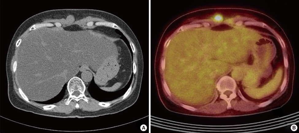
Figure 1 (A) Computed tomography scan of the abdomen, showing soft tissue density lesions in rectus abdominis muscle. (B) F-18 fluorodeoxyglucose (FDG) positron emission tomography/computed tomography, showing abdominal muscle mass with increased FDG uptake (SUVmax, 7.1).
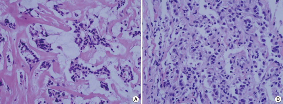
Figure 2 Diffuse scattered neoplastic cells with distraction of muscle fascicles. (A) Abdominal muscle mass (H&E stain, ×400). (B) Gluteal muscle mass (H&E stain, ×400).
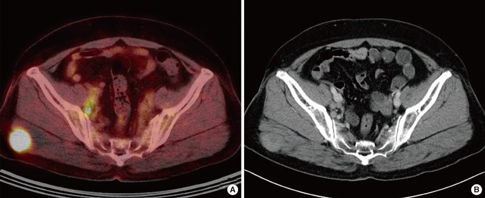
Figure 3 (A) F-18 fluorodeoxyglucose (FDG) positron emission tomography-computed tomography, showing right gluteal muscle mass with increased FDG uptake (SUVmax, 14.0). (B) Computed tomography scan of the abdomen, showing poorly-demarcated, round, isodense mass in right gluteal muscle.
Reference
-
1. Voduc KD, Cheang MC, Tyldesley S, Gelmon K, Nielsen TO, Kennecke H. Breast cancer subtypes and the risk of local and regional relapse. J Clin Oncol. 2010. 28:1684–1691.
Article2. Acinas García O, Fernández FA, Satué EG, Buelta L, Val-Bernal JF. Metastasis of malignant neoplasms to skeletal muscle. Rev Esp Oncol. 1984. 31:57–67.3. Camnasio F, Scotti C, Borri A, Fontana F, Fraschini G. Solitary psoas muscle metastasis from renal cell carcinoma. ANZ J Surg. 2010. 80:466–467.
Article4. Girard M, Kelkel E, Peoch M, Grand S, Massot C. Metastasis in the psoas muscle disclosing breast carcinoma. Ann Med Interne (Paris). 1992. 143:492–494.5. Molina-Garrido MJ, Guillén-Ponce C. Muscle metastasis of carcinoma. Clin Transl Oncol. 2011. 13:98–101.
Article6. Doo SW, Kim WB, Kim BK, Yang WJ, Yoon JH, Song YS, et al. Skeletal muscle metastases from urothelial cell carcinoma. Korean J Urol. 2012. 53:63–66.
Article7. Emmering J, Vogel WV, Stokkel MP. Intramuscular metastases on FDG PET-CT: a review of the literature. Nucl Med Commun. 2012. 33:117–120.8. Ogiya A, Takahashi K, Sato M, Kubo Y, Nishikawa N, Kikutani M, et al. Metastatic breast carcinoma of the abdominal wall muscle: a case report. Breast Cancer. Epub 2012 Mar 2. DOI: http://dx.doi.org/10.1007/s12282-012-0352-3.
Article9. Niikura N, Liu J, Hayashi N, Mittendorf EA, Gong Y, Palla SL, et al. Loss of human epidermal growth factor receptor 2 (HER2) expression in metastatic sites of HER2-overexpressing primary breast tumors. J Clin Oncol. 2012. 30:593–599.
Article10. Tuoheti Y, Okada K, Osanai T, Nishida J, Ehara S, Hashimoto M, et al. Skeletal muscle metastases of carcinoma: a clinicopathological study of 12 cases. Jpn J Clin Oncol. 2004. 34:210–214.
Article11. Hattori H, Nishimura H, Matsuoka H, Yamamoto K. FDG-PET demonstration of asymptomatic skeletal muscle metastasis from colorectal carcinoma. J Orthop Sci. 2008. 13:481–484.
Article12. Bhargava P, Verstovsek G, Stair M, Vollink J. Metastasis to psoas muscle detected by F-18 FDG PET-CT imaging. Clin Nucl Med. 2008. 33:723–724.
Article
- Full Text Links
-
- Actions
-
Cited
- CITED
-
- Close
- Share
-
- Similar articles
-
- Metastasis of Breast Carcinoma to Intercostal Muscle Detected by Breast MRI: A Case Report
- Breast Cancer Metastatic to Gluteus Maximus: A Case Report
- Metastases to Skeletal Muscles from Non-Small Cell Lung Cancer Demonstrated by 18F-FDG PET/CT
- Cutaneous Metastasis from Prostatic Cancer
- Radiological and Histological Clues in the Diagnosis of Solitary and Synchronous Breast Metastasis From Small Cell Lung Carcinoma
