STAT3, a Poor Survival Predicator, Is Associated with Lymph Node Metastasis from Breast Cancer
- Affiliations
-
- 1Department of Thyroid and Breast Surgery, Western China Hospital of Sichuan University, Chengdu, China. wangjinghxyy@sohu.com
Abstract
- PURPOSE
The aim of this study is to explore signal transducer and activator of transcription 3 (STAT3) expression in breast cancer and to analyze the detailed mechanism that STAT3 contributes to the progression of breast cancer.
METHODS
We retrospectively analyzed the clinicopathologic characteristics and overall survival (OS) of 140 breast cancer patients after curative surgery, and detected STAT3 expression, phosphorylated STAT3 (pSTAT3) expression, Ki-67 expression, vascular endothelial growth factor (VEGF)-C and -D expression in breast cancer tissues, and adjacent nontumor tissues. Survival analysis and relationship analysis were adopted for demonstrated the important mechanism of STAT3 contribution to progression of breast cancer.
RESULTS
STAT3 expression, pSTAT3 expression, Ki-67 expression, VEGF-C expression, and VEGF-D expression in breast cancer tissues were significantly higher than those in adjacent nontumor tissues, respectively. With survival analysis, only number of lymph node metastasis (N stage) was identified as the independent predictors of the OS of breast cancer patients. Besides, we demonstrated there was the most prominent correlation between STAT3 expression and lymph node metastasis in breast cancer tissues by using the multinominal regression method.
CONCLUSION
STAT3, a poor survival biomarker potential association with lymph node metastasis, was suitable for predication the OS of breast cancer patients after curative resection.
MeSH Terms
-
Breast
Breast Neoplasms
Humans
Lymph Nodes
Neoplasm Metastasis
Prognosis
Retrospective Studies
STAT3 Transcription Factor
Vascular Endothelial Growth Factor A
Vascular Endothelial Growth Factor C
Vascular Endothelial Growth Factor D
STAT3 Transcription Factor
Vascular Endothelial Growth Factor A
Vascular Endothelial Growth Factor C
Vascular Endothelial Growth Factor D
Figure
-
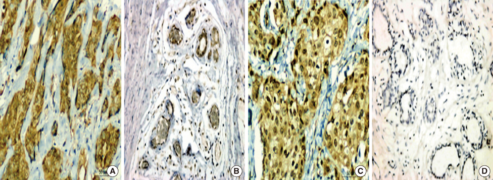
Figure 1 (A) Signal transducer and activator of transcription 3 (STAT3) expression in breast cancer tissue. (B) STAT3 expression in adjacent nontumor tissue. (C) pSTAT3 expression in breast cancer tissue. (D) pSTAT3 expression in adjacent nontumor tissue (Immunohistochemical staining, ×400).

Figure 2 (A) Vascular endothelial growth factor (VEGF)-C expression in breast cancer tissue. (B) VEGF-C expression in adjacent nontumor tissue. (C) VEGF-D expression in breast cancer tissue. (D) VEGF-D expression in adjacent nontumor tissue (Immunohistochemical staining, ×400).
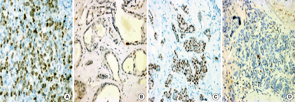
Figure 3 (A) Ki-67 expression in breast cancer tissue. (B) Ki-67 expression in adjacent nontumor tissue. (C) Estrogen receptor (ER) expression in breast cancer tissue. (D) ER expression in adjacent nontumor tissue (Immunohistochemical staining, ×400).

Figure 4 (A) Progesterone receptor (PR) expression in breast cancer tissue. (B) PR expression in adjacent nontumor tissue. (C) Human epidermal growth factor receptor 2 (HER2) expression in breast cancer tissue. (D) HER2 expression in adjacent nontumor tissue (Immunohistochemical staining, ×400).
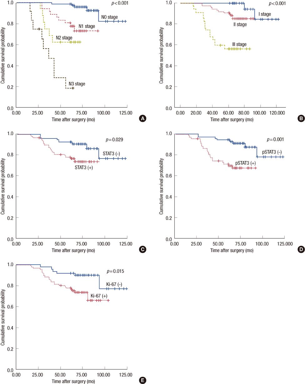
Figure 5 Survival curve for 140 breast cancer patients after curative surgery according to stage subgroup. (A) N stage (the AJCC (American Joint Committee On Cancer)) TNM classification of breast cancer). (B) TNM stage (the International Union against Cancer TNM classification of breast cancer). (C) Signal transducer and activator of transcription 3 (STAT3) expression. (D) Phosphorylated STAT3 (pSTAT3) expression. (E) Ki-67 expression.

Figure 6 Comparison mean values of lymph node metastatic counts of 140 breast cancer patients according to signal transducer and activator of transcription 3 (STAT3) expression.
Reference
-
1. Cowin P, Welch DR. Breast cancer progression: controversies and consensus in the molecular mechanisms of metastasis and EMT. J Mammary Gland Biol Neoplasia. 2007. 12:99–102.
Article2. Naik AM, Fey J, Gemignani M, Heerdt A, Montgomery L, Petrek J, et al. The risk of axillary relapse after sentinel lymph node biopsy for breast cancer is comparable with that of axillary lymph node dissection: a follow-up study of 4008 procedures. Ann Surg. 2004. 240:462–468.3. Sola M, Alberro JA, Fraile M, Santesteban P, Ramos M, Fabregas R, et al. Complete axillary lymph node dissection versus clinical follow-up in breast cancer patients with sentinel node micrometastasis: final results from the multicenter clinical trial AATRM 048/13/2000. Ann Surg Oncol. 2013. 20:120–127.
Article4. Sanuki N, Takeda A, Amemiya A, Ofuchi T, Ono M, Ogata H, et al. Outcomes of clinically node-negative breast cancer without axillary dissection: can preserved axilla be safely treated with radiation after a positive sentinel node biopsy? Clin Breast Cancer. 2013. 13:69–76.
Article5. Noguchi M, Morioka E, Ohno Y, Nakano Y, Kosaka T. The changing role of axillary lymph node dissection for breast cancer. Breast Cancer. 2013. 20:41–46.
Article6. Park J, Fey JV, Naik AM, Borgen PI, Van Zee KJ, Cody HS 3rd. A declining rate of completion axillary dissection in sentinel lymph node-positive breast cancer patients is associated with the use of a multivariate nomogram. Ann Surg. 2007. 245:462–468.
Article7. Glechner A, Wöckel A, Gartlehner G, Thaler K, Strobelberger M, Griebler U, et al. Sentinel lymph node dissection only versus complete axillary lymph node dissection in early invasive breast cancer: a systematic review and meta-analysis. Eur J Cancer. Epub 2012 Oct 17. DOI: http://dx.doi.org/10.1016/j.ejca.2012.09.010.
Article8. Mandriota SJ, Jussila L, Jeltsch M, Compagni A, Baetens D, Prevo R, et al. Vascular endothelial growth factor-C-mediated lymphangiogenesis promotes tumour metastasis. EMBO J. 2001. 20:672–682.
Article9. Van Trappen PO, Steele D, Lowe DG, Baithun S, Beasley N, Thiele W, et al. Expression of vascular endothelial growth factor (VEGF)-C and VEGF-D, and their receptor VEGFR-3, during different stages of cervical carcinogenesis. J Pathol. 2003. 201:544–554.
Article10. Couto JP, Daly L, Almeida A, Knauf JA, Fagin JA, Sobrinho-Simões M, et al. STAT3 negatively regulates thyroid tumorigenesis. Proc Natl Acad Sci U S A. 2012. 109:E2361–E2370.
Article11. Amin HM, McDonnell TJ, Ma Y, Lin Q, Fujio Y, Kunisada K, et al. Selective inhibition of STAT3 induces apoptosis and G(1) cell cycle arrest in ALK-positive anaplastic large cell lymphoma. Oncogene. 2004. 23:5426–5434.
Article12. Zhang F, Li C, Halfter H, Liu J. Delineating an oncostatin M-activated STAT3 signaling pathway that coordinates the expression of genes involved in cell cycle regulation and extracellular matrix deposition of MCF-7 cells. Oncogene. 2003. 22:894–905.
Article13. Niu G, Wright KL, Huang M, Song L, Haura E, Turkson J, et al. Constitutive Stat3 activity up-regulates VEGF expression and tumor angiogenesis. Oncogene. 2002. 21:2000–2008.
Article14. Messina JL, Yu H, Riker AI, Munster PN, Jove RL, Daud AI. Activated stat-3 in melanoma. Cancer Control. 2008. 15:196–201.
Article15. Zhao X, Sun X, Li XL. Expression and clinical significance of STAT3, P-STAT3, and VEGF-C in small cell lung cancer. Asian Pac J Cancer Prev. 2012. 13:2873–2877.
Article16. Cai QW, Li J, Li XQ, Wang JQ, Huang Y. Expression of STAT3, MMP-1 and TIMP-1 in gastric cancer and correlation with pathological features. Mol Med Rep. 2012. 5:1438–1442.17. Jiang R, Jin Z, Liu Z, Sun L, Wang L, Li K. Correlation of activated STAT3 expression with clinicopathologic features in lung adenocarcinoma and squamous cell carcinoma. Mol Diagn Ther. 2011. 15:347–352.
Article18. Deng JY, Sun D, Liu XY, Pan Y, Liang H. STAT-3 correlates with lymph node metastasis and cell survival in gastric cancer. World J Gastroenterol. 2010. 16:5380–5387.
Article19. Takemoto S, Ushijima K, Kawano K, Yamaguchi T, Terada A, Fujiyoshi N, et al. Expression of activated signal transducer and activator of transcription-3 predicts poor prognosis in cervical squamous-cell carcinoma. Br J Cancer. 2009. 101:967–972.
Article20. Fidler IJ. The pathogenesis of cancer metastasis: the 'seed and soil' hypothesis revisited. Nat Rev Cancer. 2003. 3:453–458.
Article21. Kim T, Giuliano AE, Lyman GH. Lymphatic mapping and sentinel lymph node biopsy in early-stage breast carcinoma: a metaanalysis. Cancer. 2006. 106:4–16.
Article22. Hatake K, Tokudome N, Ito Y. Next generation molecular targeted agents for breast cancer: focus on EGFR and VEGFR pathways. Breast Cancer. 2007. 14:132–149.
Article23. Yue P, Zhang X, Paladino D, Sengupta B, Ahmad S, Holloway RW, et al. Hyperactive EGF receptor, Jaks and Stat3 signaling promote enhanced colony-forming ability, motility and migration of cisplatin-resistant ovarian cancer cells. Oncogene. 2012. 31:2309–2322.
Article24. Pytowski B, Goldman J, Persaud K, Wu Y, Witte L, Hicklin DJ, et al. Complete and specific inhibition of adult lymphatic regeneration by a novel VEGFR-3 neutralizing antibody. J Natl Cancer Inst. 2005. 97:14–21.
Article25. Schoppmann SF, Jesch B, Friedrich J, Jomrich G, Maroske F, Birner P. Phosphorylation of signal transducer and activator of transcription 3 (STAT3) correlates with Her-2 status, carbonic anhydrase 9 expression and prognosis in esophageal cancer. Clin Exp Metastasis. 2012. 29:615–624.
Article26. Kim HS, Park YH, Lee J, Ahn JS, Kim J, Shim YM, et al. Clinical impact of phosphorylated signal transducer and activator of transcription 3, epidermal growth factor receptor, p53, and vascular endothelial growth factor receptor 1 expression in resected adenocarcinoma of lung by using tissue microarray. Cancer. 2010. 116:676–685.
Article27. Shah NG, Trivedi TI, Tankshali RA, Goswami JV, Jetly DH, Shukla SN, et al. Prognostic significance of molecular markers in oral squamous cell carcinoma: a multivariate analysis. Head Neck. 2009. 31:1544–1556.
Article
- Full Text Links
-
- Actions
-
Cited
- CITED
-
- Close
- Share
-
- Similar articles
-
- Correlation of tumor angiogenesis and lymph node metastasis in invasive breast carcinoma
- Prognostic value of the lymph node metastasis in patients with ampulla of Vater cancer after surgical resection
- The Prognosis of breast Cancr with more than 10 Positive Nodes
- Sentinel Lymph Node Biopsy in Breast Cancer: A Clinical Review and Update
- Sentinel Lymph Node in Breast Cancer: Review Article from a Pathologist's Point of View
