Yonsei Med J.
2016 Jul;57(4):963-967. 10.3349/ymj.2016.57.4.963.
Comparison of Predicted Total Lung Capacity and Total Lung Capacity by Computed Tomography in Lung Transplantation Candidates
- Affiliations
-
- 1Department of Radiology, Korea University Medical Center, Anam Hospital, Seoul, Korea.
- 2Department of Thoracic and Cardiovascular Surgery, Severance Hospital, Yonsei University College of Medicine, Seoul, Korea.
- 3Department of Radiology, Gangnam Severance Hospital, Yonsei University College of Medicine, Seoul, Korea.
- 4Department of Thoracic and Cardiovascular Surgery, Ajou University School of Medicine, Suwon, Korea. haamsj@aumc.ac.kr
- KMID: 2374130
- DOI: http://doi.org/10.3349/ymj.2016.57.4.963
Abstract
- PURPOSE
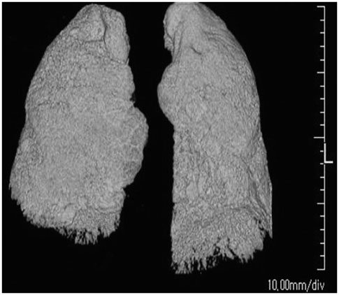
Lung size mismatch is a major cause of poor lung function and worse survival after lung transplantation (LTx). We compared predicted total lung capacity (pTLC) and TLC measured by chest computed tomography (TLC(CT)) in LTx candidates.
MATERIALS AND METHODS
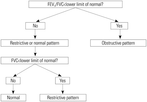
We reviewed the medical records of patients on waiting lists for LTx. According to the results of pulmonary function tests, patients were divided into an obstructive disease group and restrictive disease group. The differences between pTLC calculated using the equation of the European Respiratory Society and TLC(CT) were analyzed in each group.
RESULTS
Ninety two patients met the criteria. Thirty five patients were included in the obstructive disease group, and 57 patients were included in the restrictive disease group. pTLC in the obstructive disease group (5.50±1.07 L) and restrictive disease group (5.57±1.03 L) had no statistical significance (p=0.747), while TLC(CT) in the restrictive disease group (3.17±1.15 L) was smaller than that I the obstructive disease group (4.21±1.38 L) (p<0.0001). TLC(CT)/pTLC was 0.770 in the obstructive disease group and 0.571 in the restrictive disease group.
CONCLUSION
Regardless of pulmonary disease pattern, TLC(CT) was smaller than pTLC, and it was more apparent in restrictive lung disease. Therefore, we should consider the difference between TLC(CT) and pTLC, as well as lung disease patterns of candidates, in lung size matching for LTx.
MeSH Terms
Figure
Reference
-
1. Ouwens JP, van der Mark TW, van der Bij W, Geertsma A, de Boer WJ, Koëter GH. Size matching in lung transplantation using predicted total lung capacity. Eur Respir J. 2002; 20:1419–1422.
Article2. Eberlein M, Arnaoutakis GJ, Yarmus L, Feller-Kopman D, Dezube R, Chahla MF, et al. The effect of lung size mismatch on complications and resource utilization after bilateral lung transplantation. J Heart Lung Transplant. 2012; 31:492–500.
Article3. Eberlein M, Reed RM, Permutt S, Chahla MF, Bolukbas S, Nathan SD, et al. Parameters of donor-recipient size mismatch and survival after bilateral lung transplantation. J Heart Lung Transplant. 2012; 31:1207–1213.e7.
Article4. Mason DP, Batizy LH, Wu J, Nowicki ER, Murthy SC, McNeill AM, et al. Matching donor to recipient in lung transplantation: how much does size matter? J Thorac Cardiovasc Surg. 2009; 137:1234–1240.e1.
Article5. Massard G, Badier M, Guillot C, Reynaud M, Thomas P, Giudicelli R, et al. Lung size matching for double lung transplantation based on the submammary thoracic perimeter. Accuracy and functional results. The Joint Marseille-Montreal Lung Transplant Program. J Thorac Cardiovasc Surg. 1993; 105:9–14.
Article6. Harjula A, Baldwin JC, Starnes VA, Stinson EB, Oyer PE, Jamieson SW, et al. Proper donor selection for heart-lung transplantation. The Stanford experience. J Thorac Cardiovasc Surg. 1987; 94:874–880.7. Griffith BP, Hardesty RL, Trento A, Paradis IL, Duquesnoy RJ, Zeevi A, et al. Heart-lung transplantation: lessons learned and future hopes. Ann Thorac Surg. 1987; 43:6–16.
Article8. Goldman HI, Becklake MR. Respiratory function tests; normal values at median altitudes and the prediction of normal results. Am Rev Tuberc. 1959; 79:457–467.9. Roberts CM, MacRae KD, Winning AJ, Adams L, Seed WA. Reference values and prediction equations for normal lung function in a non-smoking white urban population. Thorax. 1991; 46:643–650.
Article10. Crapo RO, Morris AH, Clayton PD, Nixon CR. Lung volumes in healthy nonsmoking adults. Bull Eur Physiopathol Respir. 1982; 18:419–425.11. Harik-Khan RI, Fleg JL, Muller DC, Wise RA. The effect of anthropometric and socioeconomic factors on the racial difference in lung function. Am J Respir Crit Care Med. 2001; 164:1647–1654.
Article12. Bellemare JF, Cordeau MP, Leblanc P, Bellemare F. Thoracic dimensions at maximum lung inflation in normal subjects and in patients with obstructive and restrictive lung diseases. Chest. 2001; 119:376–386.
Article13. Stocks J, Quanjer PH. Reference values for residual volume, functional residual capacity and total lung capacity. ATS Workshop on Lung Volume Measurements. Official Statement of The European Respiratory Society. Eur Respir J. 1995; 8:492–506.
Article14. Knudson RJ, Slatin RC, Lebowitz MD, Burrows B. The maximal expiratory flow-volume curve. Normal standards, variability, and effects of age. Am Rev Respir Dis. 1976; 113:587–600.15. Al-Ashkar F, Mehra R, Mazzone PJ. Interpreting pulmonary function tests:recognize the pattern, and the diagnosis will follow. Cleve Clin J Med. 2003; 70:866868871–873. passim.16. Iwano S, Okada T, Satake H, Naganawa S. 3D-CT volumetry of the lung using multidetector row CT: comparison with pulmonary function tests. Acad Radiol. 2009; 16:250–256.17. Eberlein M, Permutt S, Chahla MF, Bolukbas S, Nathan SD, Shlobin OA, et al. Lung size mismatch in bilateral lung transplantation is associated with allograft function and bronchiolitis obliterans syndrome. Chest. 2012; 141:451–460.
Article18. Eberlein M, Permutt S, Brown RH, Brooker A, Chahla MF, Bolukbas S, et al. Supranormal expiratory airflow after bilateral lung transplantation is associated with improved survival. Am J Respir Crit Care Med. 2011; 183:79–87.
Article19. Aigner C, Jaksch P, Taghavi S, Wisser W, Marta G, Winkler G, et al. Donor total lung capacity predicts recipient total lung capacity after size-reduced lung transplantation. J Heart Lung Transplant. 2005; 24:2098–2102.
Article20. Santos F, Lama R, Alvarez A, Algar FJ, Quero F, Cerezo F, et al. Pulmonary tailoring and lobar transplantation to overcome size disparities in lung transplantation. Transplant Proc. 2005; 37:1526–1529.
Article21. Orens JB, Boehler A, de Perrot M, Estenne M, Glanville AR, Keshavjee S, et al. A review of lung transplant donor acceptability criteria. J Heart Lung Transplant. 2003; 22:1183–1200.
Article22. Ghio AJ, Crapo RO, Elliott CG. Reference equations used to predict pulmonary function. Survey at institutions with respiratory disease training programs in the United States and Canada. Chest. 1990; 97:400–403.
Article23. Russi EW, Karrer W, Brutsche M, Eich C, Fitting JW, Frey M, et al. Diagnosis and management of chronic obstructive pulmonary disease: the Swiss guidelines. Official guidelines of the Swiss Respiratory Society. Respiration. 2013; 85:160–174.
Article24. Irion KL, Marchiori E, Hochhegger B, Porto Nda S, Moreira Jda S, Anselmi CE, et al. CT quantification of emphysema in young subjects with no recognizable chest disease. AJR Am J Roentgenol. 2009; 192:W90–W96.
Article25. Chen F, Kubo T, Yamada T, Sato M, Aoyama A, Bando T, et al. Adaptation over a wide range of donor graft lung size discrepancies in living-donor lobar lung transplantation. Am J Transplant. 2013; 13:1336–1342.
Article26. Chen F, Kubo T, Shoji T, Fujinaga T, Bando T, Date H. Comparison of pulmonary function test and computed tomography volumetry in living lung donors. J Heart Lung Transplant. 2011; 30:572–575.
Article27. Cooper ML, Friedman PJ, Peters RM, Brimm JE. Accuracy of radiographic lung volume using new equations derived from computed tomography. Crit Care Med. 1986; 14:177–181.
Article28. Schlesinger AE, White DK, Mallory GB, Hildeboldt CF, Huddleston CB. Estimation of total lung capacity from chest radiography and chest CT in children: comparison with body plethysmography. AJR Am J Roentgenol. 1995; 165:151–154.
Article
- Full Text Links
- Actions
-
Cited
- CITED
-
- Close
- Share
- Similar articles
-
- The Effect of Lung Volume on the Size and Volume of Pulmonary Subsolid Nodules on CT: Intraindividual Comparison between Total Lung Capacity and Tidal Volume
- Comparison of Predicted Postoperative Lung Function in Pneumonectomy Using Computed Tomography and Lung Perfusion Scans
- Lung Volumes and Alveolorespiratory Function in Mitral Stenosis
- A study of radiographic determination of the total lung capacity in the normal Korean school children
- Measurement of Lung Volumes: Usefulness of Spiral CT


