Korean J Ophthalmol.
2016 Aug;30(4):258-264. 10.3341/kjo.2016.30.4.258.
Diagnostic Value of the Serum Anti-Toxocara IgG Titer for Ocular Toxocariasis in Patients with Uveitis at a Tertiary Hospital in Korea
- Affiliations
-
- 1Department of Ophthalmology, Seoul National University Bundang Hospital, Seoul National University College of Medicine, Seongnam, Korea. sejoon1@snu.ac.kr
- 2Department of Ophthalmology, Seoul National University Hospital, Seoul National University College of Medicine, Seoul, Korea.
- 3Department of Ophthalmology, Hanyang University Hospital, Seoul, Korea.
- KMID: 2373985
- DOI: http://doi.org/10.3341/kjo.2016.30.4.258
Abstract
- PURPOSE
This study evaluated the prevalence of ocular toxocariasis (OT) in patients with uveitis of unknown etiology who visited a tertiary hospital in South Korea and assessed the success of serum anti-Toxocara immunoglobulin G (IgG) enzyme-linked immunosorbent assay (ELISA) as a diagnostic test for OT.
METHODS
The records of consecutive patients with intraocular inflammation of unknown etiology were reviewed. All participants underwent clinical and laboratory investigations, including ELISA for serum anti-Toxocara IgG. OT was diagnosed based on typical clinical findings. Clinical characteristics, seropositivity, and IgG titers were compared between patients diagnosed with OT and non-OT uveitis. The seropositivity and the diagnostic value of anti-Toxocara IgG was investigated among patients with different types of uveitis.
RESULTS
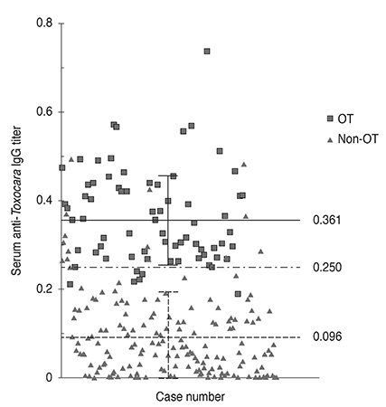
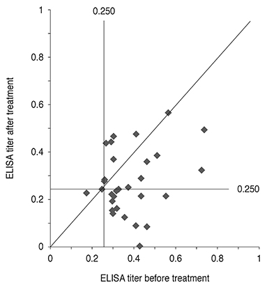
Of 238 patients with uveitis of unknown etiology, 71 (29.8%) were diagnosed with OT, and 80 (33.6%) had positive ELISA results for serum anti-Toxocara IgG. The sensitivity and specificity of the ELISA test were 91.5% (65 / 71) and 91.0% (152 / 167), respectively. The positive predictive value of the serum anti-Toxocara IgG assay was 81.3%. Among patients with anterior, intermediate, posterior, and panuveitis, the prevalence rates of OT were 8.3%, 47.1%, 44.8%, and 7.1%, respectively; the seropositivity percentages were 18.1%, 47.1%, 43.7%, and 17.9%; and the positive predictive values were 38.5%, 95.8%, 92.1%, and 40.0%. The serum anti-Toxocara IgG titer also significantly decreased following albendazole treatment.
CONCLUSIONS
OT is a common cause of intraocular inflammation in the tertiary hospital setting. Considering that OT is more prevalent in intermediate and posterior uveitis, and that the positive predictive value of the anti-Toxocara IgG assay is high, a routine test for anti-Toxocara IgG might be necessary for Korean patients with intermediate and posterior uveitis.
MeSH Terms
-
Adolescent
Adult
Aged
Aged, 80 and over
Animals
Antibodies, Anti-Idiotypic/*blood
Aqueous Humor/parasitology
Child
Enzyme-Linked Immunosorbent Assay
Eye Infections, Parasitic/*diagnosis/epidemiology/parasitology
Female
Follow-Up Studies
Humans
Immunoglobulin G/blood/*immunology
Incidence
Male
Middle Aged
Republic of Korea/epidemiology
Retrospective Studies
*Tertiary Care Centers
Toxocara canis/*immunology/isolation & purification
Toxocariasis
Uveitis/*diagnosis/epidemiology/parasitology
Young Adult
Antibodies, Anti-Idiotypic
Immunoglobulin G
Figure
Reference
-
1. Kwon SI, Lee JP, Park SP, et al. Ocular toxocariasis in Korea. Jpn J Ophthalmol. 2011; 55:143–147.2. Despommier D. Toxocariasis: clinical aspects, epidemiology, medical ecology, and molecular aspects. Clin Microbiol Rev. 2003; 16:265–272.3. Taylor MR. The epidemiology of ocular toxocariasis. J Helminthol. 2001; 75:109–118.4. Ahn SJ, Ryoo NK, Woo SJ. Ocular toxocariasis: clinical features, diagnosis, treatment, and prevention. Asia Pac Allergy. 2014; 4:134–141.5. Ahn SJ, Woo SJ, Jin Y, et al. Clinical features and course of ocular toxocariasis in adults. PLoS Negl Trop Dis. 2014; 8:e2938.6. Jin Y, Shen C, Huh S, et al. Serodiagnosis of toxocariasis by ELISA using crude antigen of Toxocara canis larvae. Korean J Parasitol. 2013; 51:433–439.7. Rubinsky-Elefant G, Hirata CE, Yamamoto JH, Ferreira MU. Human toxocariasis: diagnosis, worldwide seroprevalences and clinical expression of the systemic and ocular forms. Ann Trop Med Parasitol. 2010; 104:3–23.8. Hotez PJ, Wilkins PP. Toxocariasis: America's most common neglected infection of poverty and a helminthiasis of global importance? PLoS Negl Trop Dis. 2009; 3:e400.9. Stewart JM, Cubillan LD, Cunningham ET Jr. Prevalence, clinical features, and causes of vision loss among patients with ocular toxocariasis. Retina. 2005; 25:1005–1013.10. de Visser L, Rothova A, de Boer JH, et al. Diagnosis of ocular toxocariasis by establishing intraocular antibody production. Am J Ophthalmol. 2008; 145:369–374.11. Yoshida M, Shirao Y, Asai H, et al. A retrospective study of ocular toxocariasis in Japan: correlation with antibody prevalence and ophthalmological findings of patients with uveitis. J Helminthol. 1999; 73:357–361.12. Lim SJ, Lee SE, Kim SH, et al. Prevalence of Toxoplasma gondii and Toxocara canis among patients with uveitis. Ocul Immunol Inflamm. 2014; 22:360–366.13. Foster CS. Ocular toxocariasis. In : Foster CS, Vitale AT, editors. Diagnosis and treatment of uveitis. 2nd ed. New Delhi: JayPee Brothers Medical;2013. p. 611.
- Full Text Links
- Actions
-
Cited
- CITED
-
- Close
- Share
- Similar articles
-
- A case of presumed ocular toxocariasis in a 28-year old woman
- Atypical Form of Unilateral Optic Neuropathy Caused by Ocular Toxocariasis
- The Clinical Characteristics of Ocular Toxocariasis in Jeju Island Using Ultra-wide-field Fundus Photography
- A Case of Eosinophilic Pleural Effusion Caused by Toxocariasis without Serum Eosinophilia
- A Case Report of Intravitreal Dexamethasone Implant with Exudative Retinal Detachment for Ocular Toxocariasis Treatment


