Korean J Ophthalmol.
2016 Jun;30(3):198-205. 10.3341/kjo.2016.30.3.198.
Optical Coherence Tomography-based Diagnosis of Polypoidal Choroidal Vasculopathy in Korean Patients
- Affiliations
-
- 1Department of Ophthalmology, Konyang University College of Medicine, Daejeon, Korea.
- 2Department of Ophthalmology, Kim's Eye Hospital, Konyang University College of Medicine, Seoul, Korea. kimoph@gmail.com
- KMID: 2373975
- DOI: http://doi.org/10.3341/kjo.2016.30.3.198
Abstract
- PURPOSE
To evaluate the efficacy of an optical coherence tomography (OCT)-based diagnosis of polypoidal choroidal vasculopathy (PCV) in Korean patients.
METHODS
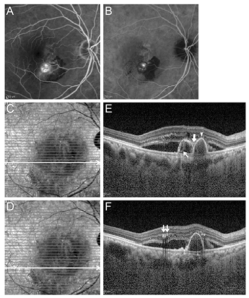
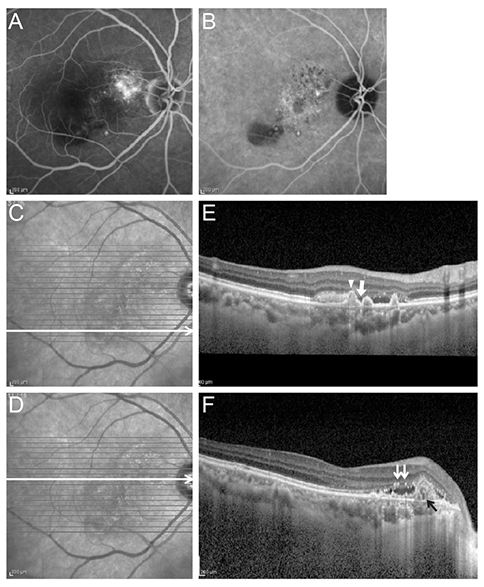
This retrospective, observational case series included 263 eyes of 263 patients (147 eyes with PCV and 116 eyes with typical exudative, age-related macular degeneration [AMD]) who had been diagnosed with treatment naïve exudative AMD. Eyes with three or more of the following OCT findings were diagnosed with PCV: multiple retinal pigment epithelial detachment (RPED), a sharp RPED peak, an RPED notch, a hyporeflective lumen representing polyps, and hyperreflective intraretinal hard exudates. The OCT-based diagnosis was compared with the gold-standard indocyanine green angiography-based method. The sensitivity and specificity of the OCT-based diagnosis was also estimated. An additional analysis was performed using a choroidal thickness criterion. Eyes with a subfoveal choroidal thickness greater than 300 µm were also diagnosed with PCV despite having only two OCT features.
RESULTS
In eyes with PCV, three or more OCT features were observed in 126 of 147 eyes (85.7%), and the incidence of typical exudative AMD was 16 of 116 eyes (13.8%). The sensitivity and specificity of an OCT-based diagnosis were 85.7% and 86.2%, respectively. After applying the choroidal thickness criterion, the sensitivity increased from 85.7% to 89.8%, and the specificity decreased from 86.2% to 84.5%.
CONCLUSIONS
The OCT-based diagnosis of PCV showed a high sensitivity and specificity in Korean patients. The addition of a choroidal thickness criterion improved the sensitivity of the method with a minimal decrease in its specificity.
Keyword
MeSH Terms
-
Aged
Choroid/blood supply/*diagnostic imaging
Choroid Diseases/*diagnosis/epidemiology/physiopathology
Diagnosis, Differential
Female
Fluorescein Angiography
Follow-Up Studies
Fundus Oculi
Humans
Incidence
Male
Republic of Korea/epidemiology
Retrospective Studies
Tomography, Optical Coherence/*methods
Visual Acuity
Figure
Reference
-
1. Yannuzzi LA, Sorenson J, Spaide RF, Lipson B. Idiopathic polypoidal choroidal vasculopathy (IPCV). Retina. 1990; 10:1–8.2. Spaide RF, Yannuzzi LA, Slakter JS, et al. Indocyanine green videoangiography of idiopathic polypoidal choroidal vasculopathy. Retina. 1995; 15:100–110.3. Koh AH, Chen LJ, Chen SJ, et al. Polypoidal choroidal vasculopathy: evidence-based guidelines for clinical diagnosis and treatment. Retina. 2013; 33:686–716.4. Martin DF, Maguire MG, Ying GS, et al. Ranibizumab and bevacizumab for neovascular age-related macular degeneration. N Engl J Med. 2011; 364:1897–1908.5. Rosenfeld PJ, Brown DM, Heier JS, et al. Ranibizumab for neovascular age-related macular degeneration. N Engl J Med. 2006; 355:1419–1431.6. Heier JS, Brown DM, Chong V, et al. Intravitreal aflibercept (VEGF trap-eye) in wet age-related macular degeneration. Ophthalmology. 2012; 119:2537–2548.7. De Salvo G, Vaz-Pereira S, Keane PA, et al. Sensitivity and specificity of spectral-domain optical coherence tomography in detecting idiopathic polypoidal choroidal vasculopathy. Am J Ophthalmol. 2014; 158:1228–1238.e1.8. Coscas G, Yamashiro K, Coscas F, et al. Comparison of exudative age-related macular degeneration subtypes in Japanese and French Patients: multicenter diagnosis with multimodal imaging. Am J Ophthalmol. 2014; 158:309–318.e2.9. Imamura Y, Engelbert M, Iida T, et al. Polypoidal choroidal vasculopathy: a review. Surv Ophthalmol. 2010; 55:501–515.10. Spaide RF, Koizumi H, Pozzoni MC. Enhanced depth imaging spectral-domain optical coherence tomography. Am J Ophthalmol. 2008; 146:496–500.11. Lee SY, Kim JG, Joe SG, et al. The therapeutic effects of bevacizumab in patients with polypoidal choroidal vasculopathy. Korean J Ophthalmol. 2008; 22:92–99.12. Oishi A, Kojima H, Mandai M, et al. Comparison of the effect of ranibizumab and verteporfin for polypoidal choroidal vasculopathy: 12-month LAPTOP study results. Am J Ophthalmol. 2013; 156:644–651.13. Koh A, Lee WK, Chen LJ, et al. EVEREST study: efficacy and safety of verteporfin photodynamic therapy in combination with ranibizumab or alone versus ranibizumab monotherapy in patients with symptomatic macular polypoidal choroidal vasculopathy. Retina. 2012; 32:1453–1464.14. Cho M, Barbazetto IA, Freund KB. Refractory neovascular age-related macular degeneration secondary to polypoidal choroidal vasculopathy. Am J Ophthalmol. 2009; 148:70–78.e1.15. Wakabayashi T, Gomi F, Sawa M, et al. Intravitreal bevacizumab for exudative branching vascular networks in polypoidal choroidal vasculopathy. Br J Ophthalmol. 2012; 96:394–399.16. Brown DM, Kaiser PK, Michels M, et al. Ranibizumab versus verteporfin for neovascular age-related macular degeneration. N Engl J Med. 2006; 355:1432–1444.17. Saito M, Kano M, Itagaki K, et al. Switching to intravitreal aflibercept injection for polypoidal choroidal vasculopathy refractory to ranibizumab. Retina. 2014; 34:2192–2201.18. Kawashima Y, Oishi A, Tsujikawa A, et al. Effects of aflibercept for ranibizumab-resistant neovascular age-related macular degeneration and polypoidal choroidal vasculopathy. Graefes Arch Clin Exp Ophthalmol. 2015; 253:1471–1477.19. Kim SW, Oh J, Kwon SS, et al. Comparison of choroidal thickness among patients with healthy eyes, early age-related maculopathy, neovascular age-related macular degeneration, central serous chorioretinopathy, and polypoidal choroidal vasculopathy. Retina. 2011; 31:1904–1911.20. Koizumi H, Yamagishi T, Yamazaki T, et al. Subfoveal choroidal thickness in typical age-related macular degeneration and polypoidal choroidal vasculopathy. Graefes Arch Clin Exp Ophthalmol. 2011; 249:1123–1128.21. Kawamura A, Yuzawa M, Mori R, et al. Indocyanine green angiographic and optical coherence tomographic findings support classification of polypoidal choroidal vasculopathy into two types. Acta Ophthalmol. 2013; 91:e474–e481.22. Koizumi H, Yamagishi T, Yamazaki T, Kinoshita S. Relationship between clinical characteristics of polypoidal choroidal vasculopathy and choroidal vascular hyperpermeability. Am J Ophthalmol. 2013; 155:305–313.e1.
- Full Text Links
- Actions
-
Cited
- CITED
-
- Close
- Share
- Similar articles
-
- Novel Findings of Polypoidal Choroidal Vasculopathy via Optical Coherence Tomography Angiography
- Diagnosing Polypoidal Choroidal Vasculopathy Using Color Fundus Photography, Optical Coherence Tomography, and Optical Coherence Tomography Angiography
- Optical Coherence Tomography of Idiopathic Polypoidal Choroidal Vasculopathy
- Availability of Optical Coherence Tomography in Diagnosis and Classification of Choroidal Neovascularization
- Epidemiologic and Clinical Characteristics of Polypoidal Choroidal Vasculopathy in Korean Patients


