MRI Findings of Uterine Tumor Resembling Ovarian Sex-Cord Tumor: A Case Report
- Affiliations
-
- 1Department of Radiology, Eulji University Hospital, Eulji University School of Medicine, Daejeon, Korea. kimhjmd@eulji.ac.kr
- 2Department of Obstetrics & Gynecology, Eulji University Hospital, Eulji University School of Medicine, Daejeon, Korea.
- 3Department of Pathology, Eulji University Hospital, Eulji University School of Medicine, Daejeon, Korea.
- KMID: 2373969
- DOI: http://doi.org/10.3348/jksr.2017.76.4.298
Abstract
- Uterine tumor resembling ovarian sex-cord tumor is a very rare uterine neoplasm that was first described by Clement and Scully in 1976. Since then, approximately 70 cases have been reported. However, these case reports have mainly described and discussed the pathologic and clinical features, and few radiologic findings have been presented. We experienced a case of a uterine tumor resembling ovarian sex-cord tumor, which was considered a uterine leiomyoma or leiomyosarcoma upon initial impression at preoperative evaluation including transvaginal ultrasonography and pelvic magnetic resonance imaging. Its diagnosis was pathologically confirmed after total abdominal hysterectomy.
MeSH Terms
Figure
-
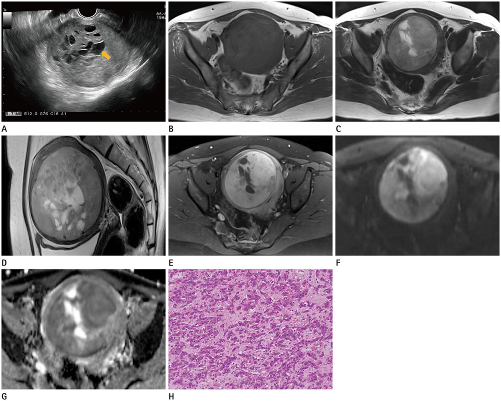
Fig. 1 Uterine tumor resembling ovarian sex-cord tumor in a 50-year-old woman. Trans-vaginal ultrasonography (A) shows an approximately 8 × 7 cm, round, solid mass with internal multi-loculated anechoic cystic portions (arrow) in the anterior wall of the uterus. Pelvic magnetic resonance imaging (B-G) shows an approximately 8.7 cm, round, solid mass in the right anterolateral wall of the uterus. On T1-weighted image (B), the mass shows iso-signal intensity compared to the myometrium. Its solid portion shows high signal intensity and its cystic portion shows bright signal intensity on T2-weighted images (C, D). Gadolinium-enhanced T1-weighted images (E) show mild enhancement except for the cystic portions. There is slight diffusion restriction in the solid portion of the mass on diffusion-weighted images (F, b = 1000) and the ADC map (G). Microscopic findings (H) of a uterine tumor resembling ovarian sex-cord tumor. Microfollicular, macrofollicular, and glandular architectural patterns are identified (hematoxylin & eosin stain, ×200).
Reference
-
1. Clement PB, Scully RE. Uterine tumors resembling ovarian sex-cord tumors. A clinicopathologic analysis of fourteen cases. Am J Clin Pathol. 1976; 66:512–552.2. Franco A, Aquino NM, Malik SL, Navarro C. Sonographic presentation of uterine sex cord-stromal tumor. J Clin Ultrasound. 1999; 27:199–201.3. Suzuki C, Matsumoto T, Fukunaga M, Itoga T, Furugen Y, Kurosaki Y, et al. Uterine tumors resembling ovarian sexcord tumors producing parathyroid hormone-related protein of the uterine cervix. Pathol Int. 2002; 52:164–168.4. Okada S, Uchiyama F, Ohaki Y, Kamoi S, Kawamura T, Kumazaki T. MRI findings of a case of uterine tumor resembling ovarian sex-cord tumors coexisting with endometrial adenoacanthoma. Radiat Med. 2001; 19:151–153.5. Morehead RP, Bowman MC. Heterologous mesodermal tumors of the uterus: report of a neoplasm resembling a granulosa cell tumor. Am J Pathol. 1945; 21:53–61.6. Hashmi AA, Faridi N, Edhi MM, Khan M. Uterine tumor resembling ovarian sex cord tumor (UTROSCT), case report with literature review. Int Arch Med. 2014; 7:47.7. Hermsen B, Bogliatto F, Bleeker M, Leidi L, Trum H, Comello E, et al. Uterine tumour resembling ovarian sex cord tumour (UTROSCT): experience with a rare disease Two case reports and overview of the literature. Obstet Gynaecol Cases Rev. 2015; 2:4.8. Calisir C, Inan U, Yavas US, Isiksoy S, Kaya T. Mazabraud's syndrome coexisting with a uterine tumor resembling an ovarian sex cord tumor (UTROSCT): a case report. Korean J Radiol. 2007; 8:438–442.9. Murase E, Siegelman ES, Outwater EK, Perez-Jaffe LA, Tureck RW. Uterine leiomyomas: histopathologic features, MR imaging findings, differential diagnosis, and treatment. Radiographics. 1999; 19:1179–1197.10. Tamai K, Koyama T, Saga T, Morisawa N, Fujimoto K, Mikami Y, et al. The utility of diffusion-weighted MR imaging for differentiating uterine sarcomas from benign leiomyomas. Eur Radiol. 2008; 18:723–730.
- Full Text Links
-
- Actions
-
Cited
- CITED
-
- Close
- Share
-
- Similar articles
-
- Uterine Tumor Resembling Ovarian Sex-Cord Tumor: A case report
- A case of uterine tumor resembling ovarian sex-cord tumor with clinical review
- A case of uterine tumor resembling ovarian sex-cord tumor
- Uterine Tumor Resembling Ovarian Sex-Cord Tumor: A case report
- Uterine Tumor Resembling Ovarian Sex-Cord Tumor: A Case Report of the Cytologic Finding
