Cavernous Hemangioma Concurrently Involving the Anterior and Middle Mediastinum and the Lung Parenchyma: A Case Report
- Affiliations
-
- 1Department of Radiology, Chungbuk National University Hospital, Cheongju, Korea. originalcrystal@hanmail.net
- 2Department of Radiology, College of Medicine and Medical Research Institute, Chungbuk National University, Cheongju, Korea.
- KMID: 2373964
- DOI: http://doi.org/10.3348/jksr.2017.76.4.273
Abstract
- Hemangioma is rarely found in the mediastinum or lung. In the mediastinum, this tumor is usually located in the anterior mediastinum and manifests as a nonspecific soft tissue mass. In the lung, it usually presents as a well-defined nodule. To the best of our knowledge, there is no case of cavernous hemangioma concurrently involving the mediastinum and lung parenchyma, except for one case of concurrent cardiac and pulmonary hemangiomas. Here, we present an interesting case of cystic anterior and middle mediastinal masses together with multiple pulmonary nodules and ground glass opacities, which were diagnosed as cavernous hemangiomas. When similar findings are encountered, clinicians should consider hemangioma in the differential diagnosis.
MeSH Terms
Figure
-
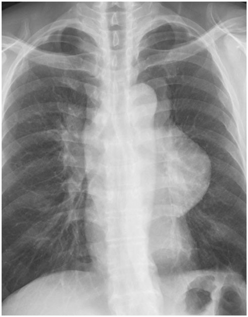
Fig. 1 A chest radiograph (postero-anterior view) shows a massive mediastinal mass and a prominent right hilum.
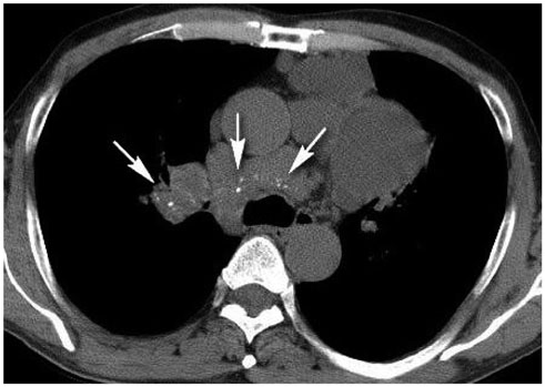
Fig. 2 An unenhanced computed tomography scan shows homogeneously attenuated thin-walled multiloculated cystic masses in the anterior mediastinum. In the middle mediastinum, there is a mass with a cystic and soft tissue density with punctate calcifications (arrows), which are thought to be phleboliths.
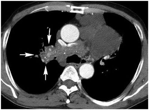
Fig. 3 A contrast-enhanced computed tomography scan obtained at the same level as that shown in Fig. 2. None of the lesions in the anterior and middle mediastinum show significant enhancement (less than 10 Hounsfield unit). The soft tissue density portion of the middle mediastinal mass is contiguous with masses in the right hilum and is located along the bronchovascular bundle in the right upper lobe (arrows).

Fig. 4 A chest computed tomography scan with a lung window setting shows multiple ill-defined ground glass opacities and multiple nodules with ground glass opacity (arrowheads) in the right upper lobe adjacent to the masses along the bronchovascular bundle, which are contiguous with those in the right hilum and the middle mediastinum. Based on histopathological examination of biopsy specimens, the diagnosis is cavernous hemangioma.
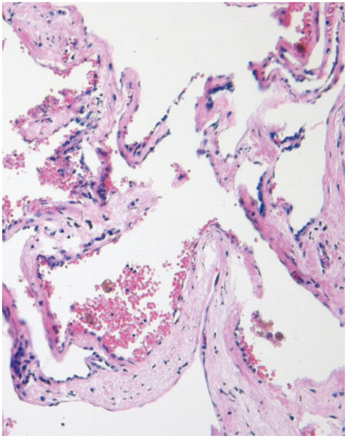
Fig. 5 Photomicrograph of the mass in the RUL shows thin-walled cavities, formed by cystic dilatation of vascular lumens. The cavities share a single wall and red blood cells are present, consistent with cavernous hemangioma (hematoxylin-eosin stain; original magnification × 100). RUL = right upper lobe
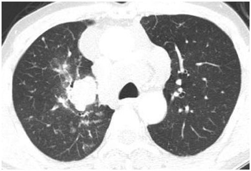
Fig. 6 A chest computed tomography scan with a lung window setting performed 4 years later. The number of mediastinal masses had decreased, probably due to the previous resection, and the stagnated multiple lung nodules and GGA remained unchanged. GGA = ground glass appearance
Reference
-
1. Weissferdt A, Moran CA. Primary vascular tumors of the lungs: a review. Ann Diagn Pathol. 2010; 14:296–308.2. Ceyhan M, Elmali M, Yildiz L. Mediastinal hemangioma and accompanying aortic arch anomaly. Pediatr Cardiol. 2008; 29:867–869.3. Agarwal PP, Seely JM, Matzinger FR. Case 130: mediastinal hemangioma. Radiology. 2008; 246:634–637.4. Cheung YC, Ng SH, Wan YL, Tan CF, Wong HF, Ng KK. Dynamic CT features of mediastinal hemangioma: more information for evaluation. Clin Imaging. 2000; 24:276–278.5. Deepak J, Babu MN, Gowrishankar BC, Ramesh S. Mediastinal hemangioma: masquerading as pleural effusion. J Indian Assoc Pediatr Surg. 2013; 18:162–164.6. Hammoumi MM, Sinaa M, Arsalane A, Oueriachi FE, Kabiri el H. Mediastinal cystic haemangiomas: a two cases report and review of the literature. Heart Lung Circ. 2014; 23:e118–e121.7. Choi SJ, Kim GY, Park CH, Lee JI, An CH. Venous hemangioma presenting as a mediastinal cyst on CT. J Korean Radiol Soc. 2007; 56:483–486.8. Kubokura H, Okamoto J, Hoshina H, Ishii H, Koizumi K, Shimizu K. Mediastinal cystic hemangioma presenting as bilateral bloody pleural effusion: a case report. J Nippon Med Sch. 2012; 79:381–384.9. Miyamoto U, Tominaga M, Tomimitsu S, Nakanishi K, Hayashi A, Irie K. A case of multiple pulmonary cavernous hemangioma. Respirol Case Rep. 2015; 3:29–32.10. Song HB, Park JC. Posterior mediastinal hemangioma mimicking Neurogenic tumor: a case report. J Korean Soc Radiol. 2015; 73:58–61.
- Full Text Links
-
- Actions
-
Cited
- CITED
-
- Close
- Share
-
- Similar articles
-
- Mediastinal Cavernous Hemangioma Involving Whole SVC: A case report
- A Case of Mediastinal Cavernous Hemangioma
- Posterior Mediastinal Hemangioma Mimicking Neurogenic Tumor: A Case Report
- Multiple Cavernous Hemangiomas of the Posterior Mediastinum, Lung, and Liver: A Case Report
- Extracerebral Cavernous Hemangioma of the Middle Cranial Fossa: Report of 2 Cases and Review of Literature
