Slow Growing Desmoid-Type Fibromatosis of the Breast: A Case Report
- Affiliations
-
- 1Department of Radiology, Sanggye Paik Hospital, Inje University College of Medicine, Seoul, Korea. radkimjy@paik.ac.kr
- 2Department of Pathology, Sanggye Paik Hospital, Inje University College of Medicine, Seoul, Korea.
- KMID: 2373962
- DOI: http://doi.org/10.3348/jksr.2017.76.4.259
Abstract
- Fibromatosis or desmoid tumor of the breast is a rare benign entity that has no metastatic potential but has significant risk of local recurrence. Its association with previous surgical or accidental trauma and Gardner's syndrome has been reported. Awareness of this lesion is important as the diagnosis is often confused with breast carcinoma. We report a case of a 44-year-old woman who presented with a palpable mass in her left breast, close to the axilla since a few months ago. She had undergone excisional biopsy for her left breast mass 15 months ago, and the diagnosis was confirmed as intraductal papilloma with atypical ductal hyperplasia. Subsequent ultrasound and core needle biopsy revealed stromal fibrosis. After 9 months, the mass showed an increase in its size and the anteroposterior to width ratio on ultrasonography compared to the previous examination, and final excisional biopsy confirmed the diagnosis of desmoid-type fibromatosis.
MeSH Terms
Figure
-
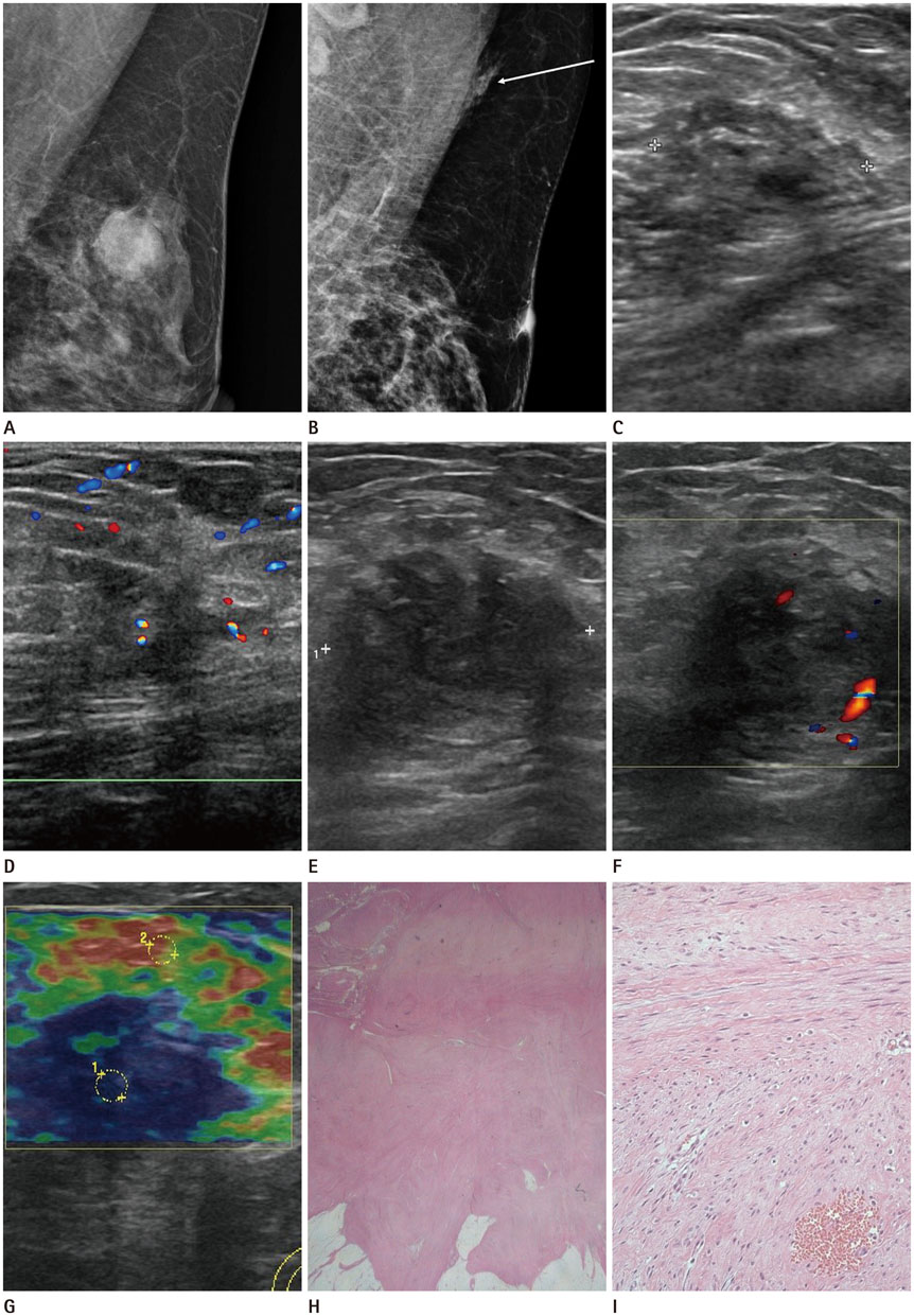
Fig. 1 Slow growing desmoid-type fibromatosis of the breast in a 44-year-old woman. A. Initial left mammogram shows a 1.9 cm-sized oval shaped isodense mass with obscured margin in the upper outer quadrant, and the diagnosis was confirmed as intraductal papilloma with atypical ductal hyperplasia after excisional biopsy. B. Follow-up left mammogram when the patient presented with a palpable mass in her left breast 15 months after excision shows asymmetry in the axilla (arrow). C. Ultrasonogram performed at the same time as B shows an ill-defined, irregularly shaped, heterogeneously hypoechoic mass at the palpable site. D. On color Doppler imaging, the mass shows slightly increased vascularity at the periphery. E. Follow-up ultrasound imaging performed 9 months after C shows an ill-defined, irregular, and heterogeneously hypoechoic mass which has increased in its size and the anteroposterior to width ratio compared to that in previous examination. F. The mass shows slightly increased vascularity at the periphery on color Doppler imaging. G. Ultrasound elastographic imaging demonstrates a very low strain value in the hypoechoic mass, showing an elasticity score of 4.5 based on the 5-point scoring system. H. Microscopic examination shows an ill-defined nodular mass with abundant pinkish collagen fibers and streaks infiltrating into the adjacent adipose tissue (hematoxylin and eosin stain, ×10). I. Microscopic examination shows hypocellular spindle cell proliferation arranged in interlacing fascicles (hematoxylin and eosin stain, ×200).
Reference
-
1. Erguvan-Dogan B, Dempsey PJ, Ayyar G, Gilcrease MZ. Primary desmoid tumor (extraabdominal fibromatosis) of the breast. AJR Am J Roentgenol. 2005; 185:488–489.2. Ha KY, Deleon P, Hamilton R. Breast fibromatosis mimicking breast carcinoma. Proc (Bayl Univ Med Cent). 2013; 26:22–24.3. Glazebrook KN, Reynolds CA. Mammary fibromatosis. AJR Am J Roentgenol. 2009; 193:856–860.4. Rosen PP, Ernsberger D. Mammary fibromatosis. A benign spindle-cell tumor with significant risk for local recurrence. Cancer. 1989; 63:1363–1369.5. Otero S, Moskovic EC, Strauss DC, Benson C, Miah AB, Thway K, et al. Desmoid-type fibromatosis. Clin Radiol. 2015; 70:1038–1045.6. Park JY, Ko MS, Kim HH, Shin HJ, Hwang JE, Gong GY, et al. Aggressive desmoid tumor mimicking breast cancer. J Korean Soc Radiol. 2010; 62:497–500.7. Lim SJ, Kang YH, Kim L, Cho YU, Lee JW, Kim YJ. Recurrent primary fibromatosis in the breast: a case report. J Korean Soc Radiol. 2012; 66:287–291.8. Nakazono T, Satoh T, Hamamoto T, Kudo S. Dynamic MRI of fibromatosis of the breast. AJR Am J Roentgenol. 2003; 181:1718–1719.
- Full Text Links
-
- Actions
-
Cited
- CITED
-
- Close
- Share
-
- Similar articles
-
- Desmoid-Type Fibromatosis Associated with Silicone Breast Implants
- Recurring Fibromatosis of Breast Following Tumorectomy: A Case Report
- Desmoid Tumor of the Facet Joint: A Case Report
- A Case of Nasal Desmoid Tumor
- Pancreatic Collision Tumor of Desmoid-Type Fibromatosis and Mucinous Cystic Neoplasm: A Case Report
