Korean J Gastroenterol.
2015 May;65(5):312-315. 10.4166/kjg.2015.65.5.312.
Spontaneous Neoplastic Remission of Hepatocellular Carcinoma
- Affiliations
-
- 1Department of Internal Medicine, Institute of Gastroenterology, Yonsei University College of Medicine, Seoul, Korea. drpjy@yuhs.ac
- 2Department of Surgery, Yonsei University College of Medicine, Seoul, Korea.
- 3Yonsei Liver Cancer Special Clinic, Severance Hospital, Seoul, Korea.
- KMID: 2373228
- DOI: http://doi.org/10.4166/kjg.2015.65.5.312
Abstract
- We report on a case of a 57-year-old male who underwent a curative resection for hepatocellular carcinoma (HCC) with histological confirmation of a spontaneously necrotized tumor. Initial serum AFP level was 4,778 ng/mL. A 3.7 cm hyperechoic mass in segment 6 of the liver was observed on ultrasonography and dynamic contrast-enhanced liver MRI showed a 3.7x3.1 cm sized HCC. He was scheduled to undergo curative surgical resection under the clinical diagnosis of an early stage HCC (Barcelona Clinic Liver Cancer stage A). Without treatment, the serum AFP level declined rapidly to 50 ng/mL over five weeks. He underwent curative wedge resection of segment 6 of the liver. Histology revealed complete necrosis of the mass rimmed by inflamed fibrous capsule on a background of HBV-related cirrhosis with infiltration of lymphoplasma cells. Exact pathophysiology underlying this event is unknown. Among the proposed mechanisms of spontaneous neoplastic remission of HCC, circulatory disturbance and activation of host immune response offer the most scientific explanation for the complete histologic necrosis of HCC in the resected mass seen in our patient.
MeSH Terms
-
Carcinoma, Hepatocellular/*diagnosis/diagnostic imaging/pathology
Hepatitis B/complications/diagnosis
Humans
Liver/diagnostic imaging/pathology
Liver Cirrhosis/etiology
Liver Neoplasms/*diagnosis/diagnostic imaging/pathology
Magnetic Resonance Imaging
Male
Middle Aged
Necrosis
Radiography
Remission, Spontaneous
Ultrasonography
alpha-Fetoproteins/analysis
alpha-Fetoproteins
Figure
-
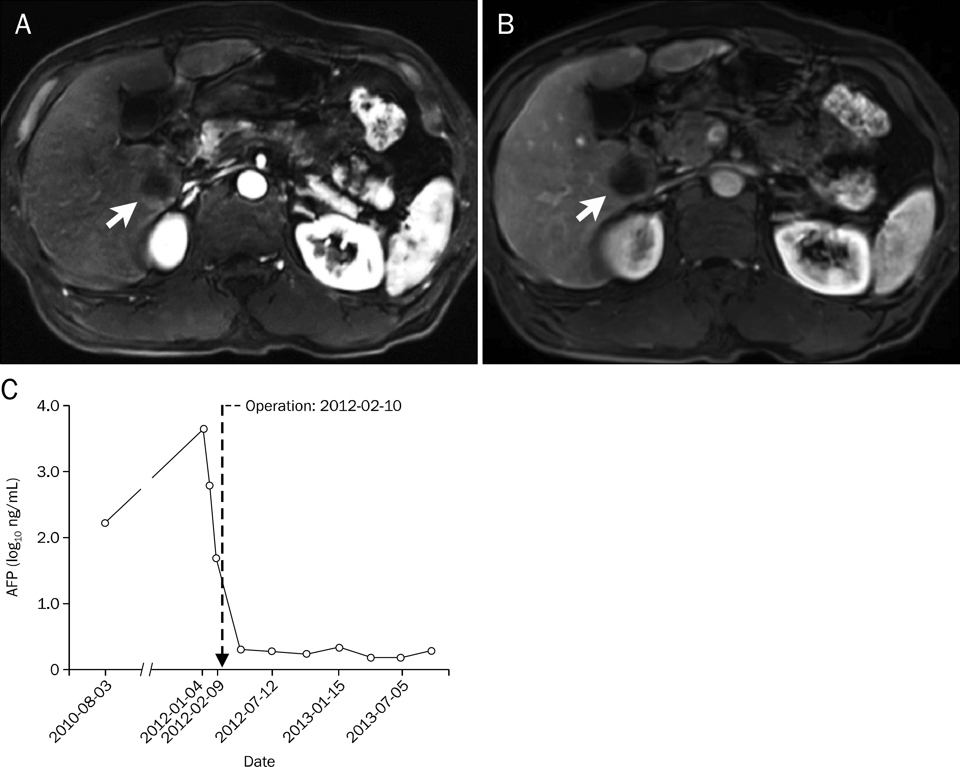
Fig. 1. Dynamic contrast-enhanced liver MRI shows a 3.7×3.1 cm mass (arrows) with arterial enhancement (A) and delayed washout (B) in T1-weighted images. (C) Graph illustrates the changes in AFP levels from referral to post resection of the tumor. Serum AFP level was 4,778 ng/mL at referral, but declined spontaneously to 650 ng/mL and dropped further to 50 ng/mL prior to surgery. After surgery, serum AFP level remained within normal limits.
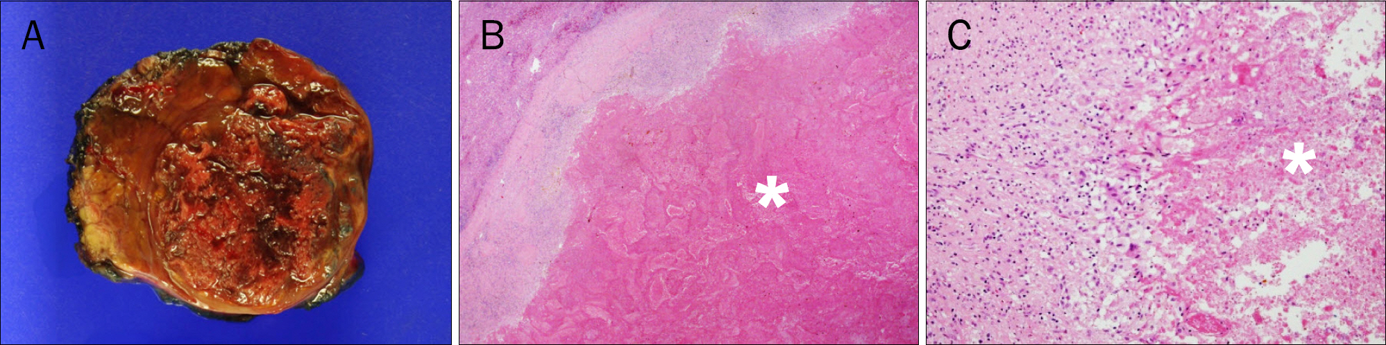
Fig. 2. (A) Gross appearance of the resected tumor shows an expanding type of mass with necrotic features. (B, C) Histology reveals complete necrosis of hepatocellular carcinoma (asterisks) rimmed by inflamed fibrous capsule with lymphoplasma cell infiltration (H&E stain; B: ×12.5, C: ×100).
Reference
-
References
1. Forner A, Llovet JM, Bruix J. Hepatocellular carcinoma. Lancet. 2012; 379:1245–1255.
Article2. Cole WH. Efforts to explain spontaneous regression of cancer. J Surg Oncol. 1981; 17:201–209.
Article3. Huz JI, Melis M, Sarpel U. Spontaneous regression of hepatocellular carcinoma is most often associated with tumour hy-poxia or a systemic inflammatory response. HPB (Oxford). 2012; 14:500–505.
Article4. Everson TC, Cole WH. Spontaneous regression of malignant disease. J Am Med Assoc. 1959; 169:1758–1759.
Article5. Sasaki T, Fukumori D, Yamamoto K, Yamamoto F, Igimi H, Yamashita Y. Management considerations for purported spontaneous regression of hepatocellular carcinoma: a case report. Case Rep Gastroenterol. 2013; 7:147–152.
Article6. Okano A, Ohana M, Kusumi F, Nabeshima M. Spontaneous regression of hepatocellular carcinoma due to disruption of the feeding artery. Case Rep Oncol. 2013; 6:180–185.
Article7. Oquiñena S, Iñarrairaegui M, Vila JJ, Alegre F, Zozaya JM, Sangro B. Spontaneous regression of hepatocellular carcinoma: three case reports and a categorized review of the literature. Dig Dis Sci. 2009; 54:1147–1153.
Article8. Ohtani H, Yamazaki O, Matsuyama M, et al. Spontaneous regression of hepatocellular carcinoma: report of a case. Surg Today. 2005; 35:1081–1086.
Article9. Iiai T, Sato Y, Nabatame N, Yamamoto S, Makino S, Hatakeyama K. Spontaneous complete regression of hepatocellular carcinoma with portal vein tumor thrombus. Hepatogastroenterology. 2003; 50:1628–1630.10. Morimoto Y, Tanaka Y, Itoh T, Yamamoto S, Mizuno H, Fushimi H. Spontaneous necrosis of hepatocellular carcinoma: a case report. Dig Surg. 2002; 19:413–418.
Article11. Stoelben E, Koch M, Hanke S, et al. Spontaneous regression of hepatocellular carcinoma confirmed by surgical specimen: report of two cases and review of the literature. Langenbecks Arch Surg. 1998; 383:447–452.
Article12. Lam KC, Ho JC, Yeung RT. Spontaneous regression of hepatocellular carcinoma: a case study. Cancer. 1982; 50:332–336.
Article13. Yoo JJ, Lee JH, Lee KB, et al. Pathologically confirmed spontaneous partial regression of hepatocellular carcinoma. Korean J Med. 2014; 86:198–203.
Article14. Park HS, Jang KY, Kim YK, Cho BH, Moon WS. Hepatocellular carcinoma with massive lymphoid infiltration: a regressing phe-nomenon? Pathol Res Pract. 2009; 205:648–652.
Article15. Wada Y, Nakashima O, Kutami R, Yamamoto O, Kojiro M. Clinicopathological study on hepatocellular carcinoma with lym-phocytic infiltration. Hepatology. 1998; 27:407–414.
Article
- Full Text Links
-
- Actions
-
Cited
- CITED
-
- Close
- Share
-
- Similar articles
-
- A Case of Complete Remission in Ruptured Hepatocellular Carcinoma after One -time Transcatheter Arterial Chemoembolization
- Spontaneous Complete Remission of Hepatocellular Carcinoma
- Kupffer Cells in Hepatocellular Carcinoma
- Arginase-1 and P-glycoprotein are downregulated in canine hepatocellular carcinoma
- A Case of Spontaneous Regression of Hepatocellular Carcinoma after Ultrasound Guided Liver Biopsy
