Solitary Pigmented Plexiform Neurofibroma in Lower Eyelid: A Case Report
- Affiliations
-
- 1Department of Ophthalmology, Chonnam National University Medical School, Gwangju, Korea. wchoi82@hanmail.net
- 2Department of Pathology, Chonnam National University Medical School, Gwangju, Korea.
- KMID: 2371915
- DOI: http://doi.org/10.3341/jkos.2017.58.3.333
Abstract
- PURPOSE
Solitary plexiform neurofibroma of the eyelid without neurofibromatosis is a rare disease. We report a case of solitary plexiform pigmented neurofibroma of the eyelid without neurofibromatosis.
CASE SUMMARY
A 12-year-old male visited our clinic with a painless palpable subcutaneous mass on the right lower eyelid. He had a history of Batter syndrome and attention deficit hyperactivity disorder. On initial presentation, clinical features regarding neurofibromatosis such as Lisch nodule, optic nerve glioma, or high myopia were not observed. We performed excision and biopsy of the lower lid mass under general anesthesia. Macroscopically, the tumor was 4.0 × 1.5 × 1.5 cm in size with irregular nodules. Microscopically, the tumor consisted of multiple, variably sized tortous enlarged nerve fascicles with clusters of pigmented cells. Immunohistochemical results revealed expression of S-100 protein. Pigmented cells express both S-100 and melan-A proteins, while nonpigmented cells express S-100 protein only. The tumor was finally diagnosed as plexiform pigmented neurofibroma. Dermatological evaluation revealed no evidence of systemic neurofibromatosis.
CONCLUSIONS
Plexiform neurofibroma should be considered in the differential diagnosis of an eyelid mass, even if the patient does not have a history or clinical features of neurofibromatosis. Plexiform neurofibroma can be successfully managed with surgical excision.
MeSH Terms
Figure
-

Figure 1. External photograph. Non-tender, palpable mass was observed on the right lower eyelid.
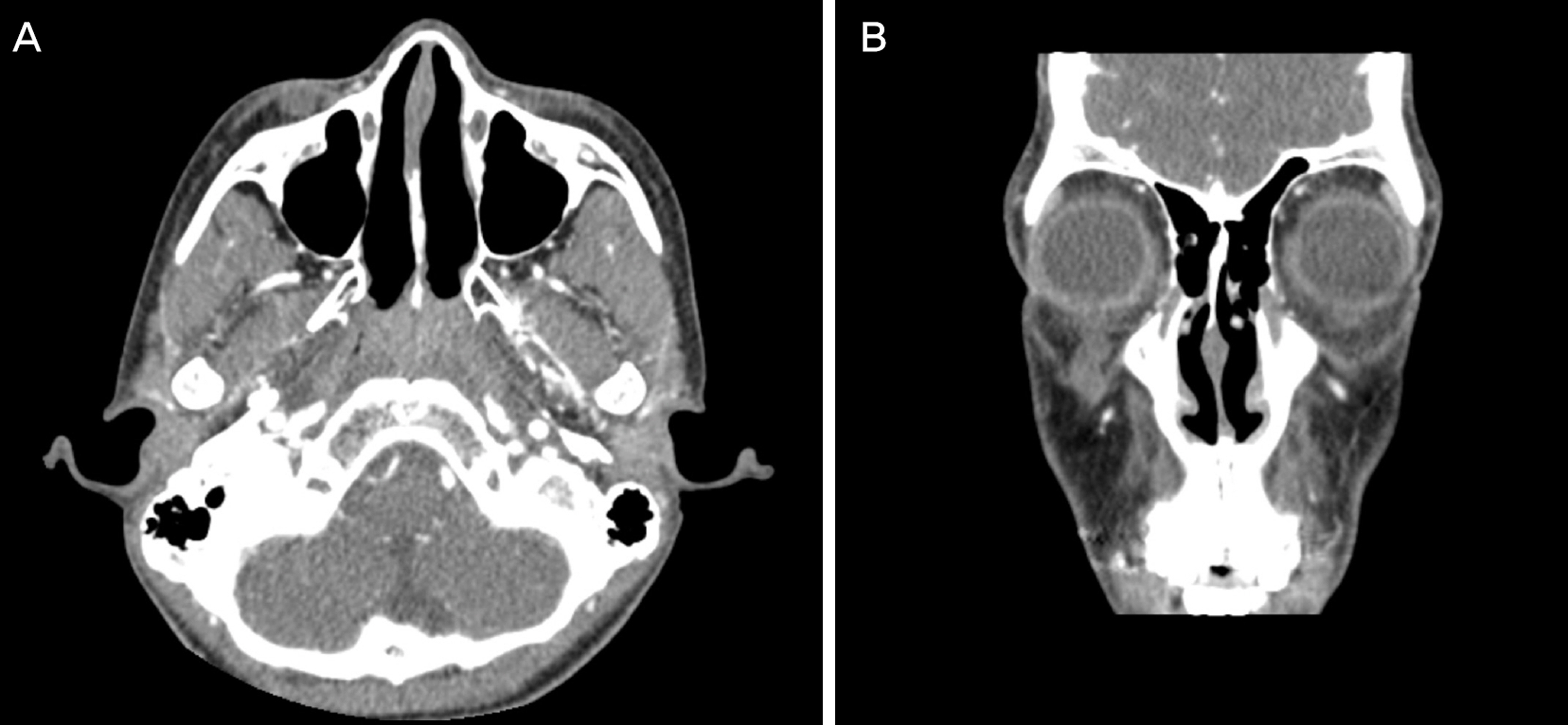
Figure 2. Enhanced Orbital computed tomography. Images showed about 3.8 × 1.5 × 1.5 cm sized non-enhanced cystic lesion at subcutaneous tissue of the right lower lid and focal skin thickening. (A) Axial view. (B) Coronal view.

Figure 3. The patient underwent surgical resection of the tumor. (A) Photomicrograph of plexiform pigmented neurofibroma (Hematoxylin-eosin, original magnification ×40). Multiple, variable sized tortuous hypertrophic nerves. (B) High power photo-micrograph of plexiform pigmented neurofibroma (Hematoxylin-eosin, original magnification ×200). Clusters of melanin pig-mented cells surrounding hypertrophied nerve of neurofibroma. (C) High power photomicrograph (S-100, original magnification ×200). Pigmented and nonpigmented tumor cells expressing S-100 protein. (D) High power photomicrograph (Melan-A, original magnification ×200) Pigmented cells with strong immunoreactivity for Melan-A, in contrast to nonpigmented cells.
Reference
-
References
1. Song BR, Yoo JH. Solitary neurofibroma in orbit. J Korean Ophthalmol Soc. 1990; 31:525–8.2. Arigon V, Binaghi M, Sabouret C. . Usefulness of systematic ophthalmologic investigations in neurofibromatosis 1: a cross-sec-tional study of 211 patients. Eur J Ophthalmol. 2002; 12:413–8.
Article3. Bechtold D, Hove HD, Prause JU. . Plexiform neurofibroma of the eye region occurring in patients without neurofibromatosis type 1. Ophthal Plast Reconstr Surg. 2012; 28:413–5.
Article4. Jung JW, Lee JH, Shyn KH, Chi MJ. A case of plexiform neuro-fibroma with severe ptosis and proptosis. J Korean Ophthalmol Soc. 2007; 48:725–30.5. Schaffer JV, Chang MW, Kovich OI. . Pigmented plexiform neurofibroma: Distinction from a large congenital melanocytic nevus. J AM Acad Dermatol. 2007; 56:862–8.
Article6. Huang W, Chong WS. Facial plexiform neurofinorma: is it truly just skin deep? BMJ Case Rep 2013. 2013; (pii):bcr2013200716.7. Shi L, Liu FJ, Jia QH. . Solitary plexiform neurofibroma of the stomach: a case report. World J Gastroenterol. 2014; 20:5153–6.
Article8. Rong AJ, Ledgerwood LG, Jin LW, Tollefson TT. Solitary plexi-form neurofibroma of the forehead: A rear and unusual presentation. Int J Pediatr Otorhinolaryngol Extra. 2014; 9:15–7.
