Giant Epidermal Inclusion Cyst in the Male Breast: A Case Report
- Affiliations
-
- 1Department of Radiology, Daejin Medical Center Bundang Jesaeng General Hospital, Seongnam, Korea. woonju@dmc.or.kr
- 2Department of General Surgery, Daejin Medical Center Bundang Jesaeng General Hospital, Seongnam, Korea.
- 3Department of Pathology, Daejin Medical Center Bundang Jesaeng General Hospital, Seongnam, Korea.
- KMID: 2371687
- DOI: http://doi.org/10.3348/jksr.2017.76.3.206
Abstract
- Giant epidermal inclusion cyst is a rare disease entity, and the occurrence of this cyst in the male breast is extremely rare. We report a case of giant epidermal inclusion cyst in the breast, which presented as a palpable and painful right breast mass in a 63-year-old man. The sonographic and computed tomography (CT) features are described in-depth. Physical examination revealed a firm, well-defined mass in the upper central portion of the right breast. Ultrasonography showed a 5.2 cm sized, oval, circumscribed, and complex cystic and solid mass with posterior acoustic enhancement, and CT showed a well-defined homogeneous low density mass without enhancement in the right breast. Surgical excision was performed, and pathological examination revealed a giant epidermal inclusion cyst.
MeSH Terms
Figure
-
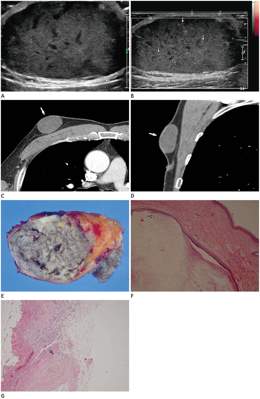
Fig. 1 A 63-year-old man with a growing palpable mass in the right breast, revealing as giant epidermal inclusion cyst. A, B. Transverse scan (A) and longitudinal color Doppler image (B) of breast sonography show an oval shaped, circumscribed, and complex cystic and solid mass in the right breast. It shows the typical pseudotestis pattern with echogenic reflectors (white arrows in B), filiform anechoic areas (black arrows in B), dermal attachment (open arrows in B) and posterior acoustic enhancement (white arrows in A) without vascularity in the mass. C, D. Axial scan (C) and sagittal scan (D) of chest CT show a well-defined homogeneous low density mass (arrows) without enhancement in the upper central portion of the right breast. E. Excised specimen showing a ruptured epidermal inclusion cyst, filled with foul-smelling, dirty greasy material. F. An intradermal cyst lined by keratinizing stratified squamous epithelium is seen. Note layers of desquamated keratin (arrow) within the cyst (hematoxylin and eosin stain × 40). G. Cholesterol clefts (arrow) surrounded by multinucleated giant cells, lymphocytes and histiocytes are noted, suggesting a microruptured cyst (hematoxylin and eosin stain × 100).
Reference
-
1. Ziadi S, Trimeche M, Hammedi F, Sriha B, Jomaa W, Mokni M, et al. Squamous cell carcinoma arising from an epidermal inclusion cyst: a case report. N Am J Med Sci. 2010; 2:46–47.2. Motabar AR. Epidermal inclusion cysts of the breast. Med J Islam Repub Iran. 2009; 22:207–211.3. Mote DG, Shukla AA. Epidermal inclusion cyst masquerading breast lump. Indian J Surg. 2011; 73:458–459.4. Rahul K, Panda A, Handa N, Hari S. Epidermal inclusion cyst in a male breast: parallel linear echoes (tram-track appearance) on sonography as a diagnostic clue. BMJ Case Rep. 2015; 12. 07. [Epub]. DOI: 10.1136/bcr-2015-213045.5. D'Orsi CJ, Sickles EA, Mendelson EB, Morris EA. ACR BIRADS Atlas®, breast imaging reporting and data system. 5th ed. Reston, VA: American College of Radiology;2013.6. Im JT, Park BY. Giant epidermal cyst on posterior scalp. Arch Plast Surg. 2013; 40:280–282.7. Lee YA, Park SG. Giant sized epidermal inclusion cyst of the breast initially mimicking a large fibroadenoma or phyllodes tumor. J Korean Surg Soc. 2012; 83:107–110.8. Kang EJ, Lee JH, Kim EK, Park Y, Jung JS, Kown H, et al. Epidermal inclusion cyst after breast reconstruction with TRAM flaps. J Korean Soc Radiol. 2010; 63:79–82.9. Huang CC, Ko SF, Huang HY, Ng SH, Lee TY, Lee YW, et al. Epidermal cysts in the superficial soft tissue: sonographic features with an emphasis on the pseudotestis pattern. J Ultrasound Med. 2011; 30:11–17.10. Yitta S, Singer CI, Toth HB, Mercado CL. Image presentation. Sonographic appearances of benign and malignant male breast disease with mammographic and pathologic correlation. J Ultrasound Med. 2010; 29:931–947.
- Full Text Links
-
- Actions
-
Cited
- CITED
-
- Close
- Share
-
- Similar articles
-
- Giant sized epidermal inclusion cyst of the breast initially mimicking a large fibroadenoma or phyllodes tumor
- A Ruptured Epidermal Inclusion Cyst in the Breast Presenting as a Recurrent Abscess
- Epidermal Inclusion Cyst after Breast Reconstruction with TRAM Flaps
- A Case of Digital Myxoid Cyst Coexisting with Epidermal Inclusion Cyst
- Giant Epidermal Cyst - A Case Repot -
