Central Retinal Artery Occlusion by Left Atrial Myxoma
- Affiliations
-
- 1Department of Ophthalmology, Kyungpook National University School of Medicine, Daegu, Korea. itkim@knu.ac.kr
- KMID: 2368684
- DOI: http://doi.org/10.3341/kjo.2017.31.1.88
Abstract
- No abstract available.
Figure
-
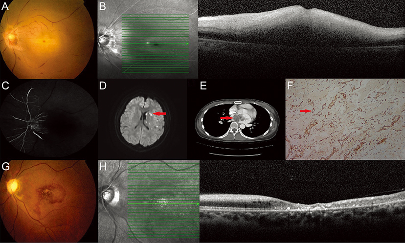
Fig. 1 (A) Fundus photograph of the left eye shows retinal artery attenuation and a cherry-red spot. (B) Optical coherence tomography shows severe edema in the macula. (C) Fluorescein angiography of the left eye shows a delay in arterial filling at 5 minutes 37 seconds. (D) Brain diffusion-weighted magnetic resonance imaging shows acute multiple signal changes (red arrow) in the left middle cerebral artery territory. (E) Contrast-enhanced chest computed tomography scan shows a heterogeneous mass (red arrow) with fluid density in the left atrium. (F) Section from excised tissue shows CD31 positivity in the abundant blood vessels (red arrow), original magnification ×100. (G,H) Follow-up fundus photograph and optical coherence tomography show atrophy of macula and optic disc.
Reference
-
1. Varma DD, Cugati S, Lee AW, Chen CS. A review of central retinal artery occlusion: clinical presentation and management. Eye (Lond). 2013; 27:688–697.2. O'Rourke F, Dean N, Mouradian MS, et al. Atrial myxoma as a cause of stroke: case report and discussion. CMAJ. 2003; 169:1049–1051.3. Pucci A, Gagliardotto P, Zanini C, et al. Histopathologic and clinical characterization of cardiac myxoma: review of 53 cases from a single institution. Am Heart J. 2000; 140:134–138.4. Rafuse PE, Nicolle DA, Hutnik CM, Pringle CE. Left atrial myxoma causing ophthalmic artery occlusion. Eye (Lond). 1997; 11(Pt 1):25–29.
- Full Text Links
-
- Actions
-
Cited
- CITED
-
- Close
- Share
-
- Similar articles
-
- Incomplete Central Retinal Artery Occlusion
- Central Retinal Artery Occlusion after Cervical Spine Surgery in Prone Position: A Case Report
- The Successful Treatment of a Case of Central Retinal Artery Occlusion
- A Case Report of Central Retinal Artery Occlusion Caused by Cardiac Myxoma
- Central Retinal Artery Occlusion Masquerading as Branch Retinal Artery Occlusion
