J Adv Prosthodont.
2017 Feb;9(1):67-73. 10.4047/jap.2017.9.1.67.
The effect of coloring liquid dipping time on the fracture load and color of zirconia ceramics
- Affiliations
-
- 1Department of Prosthodontics, Faculty of Dentistry, Cukurova University, Adana, Turkey. oekren@cu.edu.tr
- KMID: 2368214
- DOI: http://doi.org/10.4047/jap.2017.9.1.67
Abstract
- PURPOSE
The aims of the study were to evaluate the fracture load of zirconia core material after dipping in coloring liquid at different time intervals and to compare the color of dipped blocks with that of prefabricated shaded blocks.
MATERIALS AND METHODS
3-unit bridge frameworks were designed digitally. Sixty frameworks were fabricated using uncolored zirconia blocks by CAD/CAM and divided into 4 groups randomly (n = 15). Group 2 (G2) was subjected to coloring liquids for 2 minutes, Group 4 (G4) for 4 minutes, and Group 6 (G6) for 6 minutes. CFS group was not subjected to any coloring procedure. After coloring, color differences between the test groups and a prefabricated shaded zirconia group (CPZ, n = 15) were evaluated by using a spectrophotometer. Fracture test was conducted immediately after shade evaluation with a Testometric test device at a cross-head speed of 1 mm/sec. Statistical analysis for evaluating color and fracture load was performed by using one way ANOVA followed by Tukey HSD test (P ≤ .05). Weibull analysis was conducted for distribution of fracture load.
RESULTS
There was no difference in terms of fracture load and color between CFS (1176.681 N) and G2 (985.638 N) group and between CPZ (81.340) and G2 (81.140) group, respectively. Fracture load values of G4 (779.340 N) and G6 (935.491 N) groups were statistically significantly lower than that of CFS group (P ≤ .005). The color values of G4 (79.340) and G6 (79.673) groups were statistically different than that of CPZ group (P ≤ .005).
CONCLUSION
Prolonged immersion of zirconia in coloring liquid not only negatively affected the fracture load of the zirconia being tested in the current study but also deteriorated the desired shade of the restoration.
Keyword
Figure
-
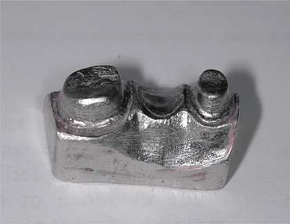
Fig. 1 Metal master model used in the study.

Fig. 2 Test Specimens prepared for G6 test group.
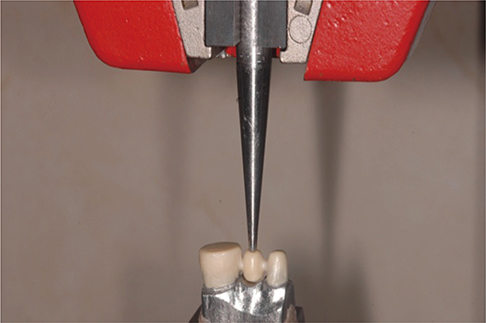
Fig. 3 Testing of specimens with Testometric. The indenter was located on the depression of the functional cusp.
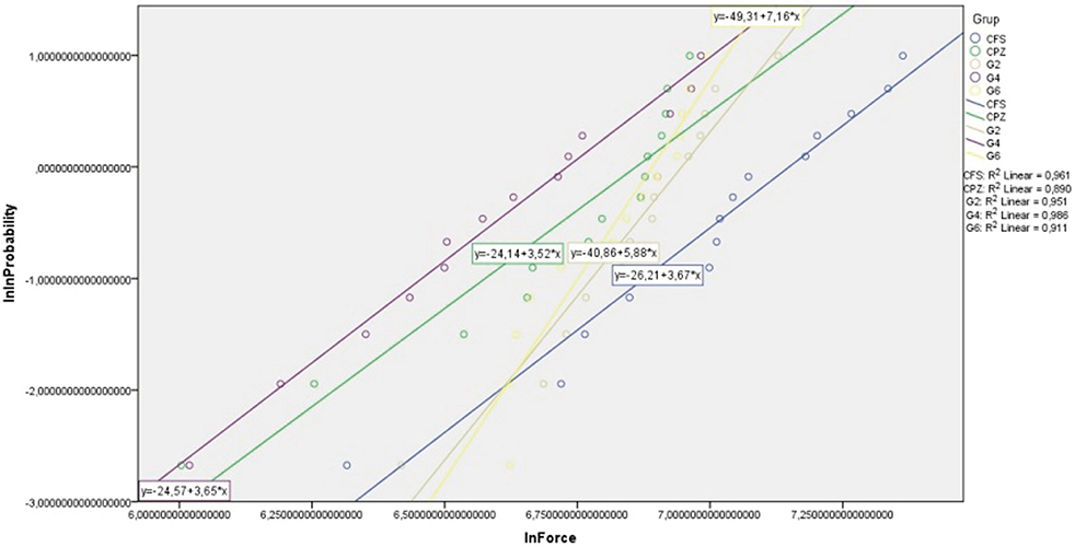
Fig. 4 Weibull graph.
Cited by 1 articles
-
Effect of different coloring liquids on the flexural strength of multilayered zirconia
Na-Kyoung Yu, Mi-Gyoung Park
J Adv Prosthodont. 2019;11(4):209-214. doi: 10.4047/jap.2019.11.4.209.
Reference
-
1. Isgrò G, Pallav P, van der Zel JM, Feilzer AJ. The influence of the veneering porcelain and different surface treatments on the biaxial flexural strength of a heat-pressed ceramic. J Prosthet Dent. 2003; 90:465–473.2. Cardelli P, Manobianco FP, Serafini N, Murmura G, Beuer F. Full-arch, Implant-supported monolithic zirconia rehabilitations: Pilot clinical evaluation of wear against natural or composite teeth. J Prosthodont. 2015; 10. 05.3. O'Brien WJ. Dental materials and their selection. 3rd ed. Chicago: Quintessence;2002. p. 210–224.4. Piconi C, Maccauro G. Zirconia as a ceramic biomaterial. Biomaterials. 1999; 20:1–25.5. Guazzato M, Albakry M, Quach L, Swain MV. Influence of surface and heat treatments on the flexural strength of a glass-infiltrated alumina/zirconia-reinforced dental ceramic. Dent Mater. 2005; 21:454–463.6. Guazzato M, Albakry M, Ringer SP, Swain MV. Strength, fracture toughness and microstructure of a selection of all-ceramic materials. Part II. Zirconia-based dental ceramics. Dent Mater. 2004; 20:449–456.7. Denry I, Kelly JR. State of the art of zirconia for dental applications. Dent Mater. 2008; 24:299–307.8. Kosmac T, Oblak C, Jevnikar P, Funduk N, Marion L. The effect of surface grinding and sandblasting on flexural strength and reliability of Y-TZP zirconia ceramic. Dent Mater. 1999; 15:426–433.9. Hannink RHJ, Kelly PM, Muddle BC. Transformation toughening in zirconia-containing ceramics. J Am Ceram Soc. 2000; 83:461–487.10. White SN, Miklus VG, McLaren EA, Lang LA, Caputo AA. Flexural strength of a layered zirconia and porcelain dental all-ceramic system. J Prosthet Dent. 2005; 94:125–131.11. Sailer I, Fehér A, Filser F, Lüthy H, Gauckler LJ, Schärer P, Franz Hämmerle CH. Prospective clinical study of zirconia posterior fixed partial dentures: 3-year follow-up. Quintessence Int. 2006; 37:685–693.12. Sailer I, Fehér A, Filser F, Gauckler LJ, Lüthy H, Hämmerle CH. Five-year clinical results of zirconia frameworks for posterior fixed partial dentures. Int J Prosthodont. 2007; 20:383–388.13. Roediger M, Gersdorff N, Huels A, Rinke S. Prospective evaluation of zirconia posterior fixed partial dentures: fouryear clinical results. Int J Prosthodont. 2010; 23:141–148.14. Lee WF, Feng SW, Lu YJ, Wu HJ, Peng PW. Effects of two surface finishes on the color of cemented and colored anatomic-contour zirconia crowns. J Prosthet Dent. 2016; 116:264–268.15. Tuncel İ, Özat P, Eroğlu E. Effects of coloring procedures on zirconia/veneer ceramics bond strength. J Adv Prosthodont. 2014; 6:451–455.16. Kim HK, Kim SH. Effect of the number of coloring liquid applications on the optical properties of monolithic zirconia. Dent Mater. 2014; 30:e229–e237.17. Hjerppe J, Närhi T, Fröberg K, Vallittu PK, Lassila LV. Effect of shading the zirconia framework on biaxial strength and surface microhardness. Acta Odontol Scand. 2008; 66:262–267.18. Sedda M, Vichi A, Carrabba M, Capperucci A, Louca C, Ferrari M. Influence of coloring procedure on flexural resistance of zirconia blocks. J Prosthet Dent. 2015; 114:98–102.19. Pittayachawan P, McDonald A, Petrie A, Knowles JC. The biaxial flexural strength and fatigue property of Lava Y-TZP dental ceramic. Dent Mater. 2007; 23:1018–1029.20. Shah K, Holloway JA, Denry IL. Effect of coloring with various metal oxides on the microstructure, color, and flexural strength of 3Y-TZP. J Biomed Mater Res B Appl Biomater. 2008; 87:329–337.21. Ardlin BI. Transformation-toughened zirconia for dental inlays, crowns and bridges: chemical stability and effect of low-temperature aging on flexural strength and surface structure. Dent Mater. 2002; 18:590–595.22. Wen N, Yi Y, Zhang W, Liu H, Tian J. Properties of dental alumina ceramics infiltrated with colored glass. Rare Metal Mater Eng. 2007; 36:82–84.23. Shetty DK, Rosenfield AR, McGuire P, Duckworth WH. Biaxial flexure test for ceramics. American Ceram Soc Bulletin. 1980; 59:1193–1197.24. Mijoska A, Popovska M. Evaluation of different in vitro testing methods for mechanical properties of veneer ceramics. Pril (Makedon Akad Nauk Umet Odd Med Nauki). 2015; 36:225–230.25. Gargari M, Gloria F, Cappello A, Ottria L. Strength of zirconia fixed partial dentures: review of the literature. Oral Implantol (Rome). 2010; 3:15–24.26. Studart AR, Filser F, Kocher P, Gauckler LJ. In vitro lifetime of dental ceramics under cyclic loading in water. Biomaterials. 2007; 28:2695–2705.27. Quinn JB, Quinn GD. A practical and systematic review of Weibull statistics for reporting strengths of dental materials. Dent Mater. 2010; 26:135–147.
- Full Text Links
-
- Actions
-
Cited
- CITED
-
- Close
- Share
-
- Similar articles
-
- Effect of coloring liquids on biaxial flexural strength of monolithic zirconia
- Effect of different coloring liquids on the flexural strength of multilayered zirconia
- The effect of repeated firings on the color of zirconia-based all-ceramic system
- Effect of the amount of thickness reduction on color and translucency of dental monolithic zirconia ceramics
- Prosthetic treatment in esthetic area with monolithic zirconia using coloring liquid: a case report
