Ann Dermatol.
2014 Jun;26(3):428-430.
Intramuscular Vascular Malformation of the Temporalis Muscle: A Case Report and Review of the Literature
- Affiliations
-
- 1Department of Dermatology, Kyung Hee University School of Medicine, Seoul, Korea. bellotte@hanmail.net
Abstract
- No abstract available.
MeSH Terms
Figure
-
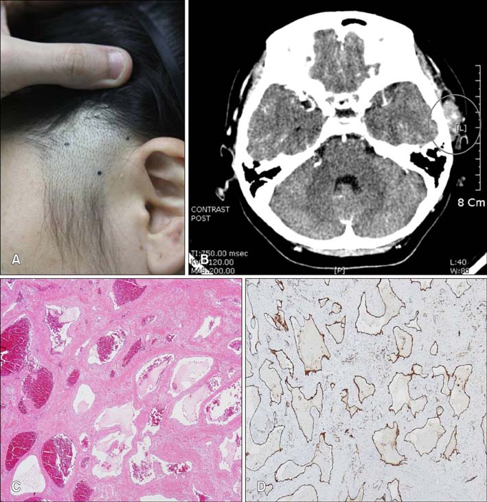
Fig. 1 (A) A 2×2 cm sized, soft, movable and nontender mass on the left temporal area. (B) A 2 cm sized mass in the left temporalis muscle, the mass is slightly and homogeneously enhanced with radio-contrast dye (contrast enhanced computed tomography scan). (C) Numerous dilated, rather thin walled vessels filled with serous fluid and red blood cells are seen (H&E, ×100). (D) Vascular channels and flattened endothelium are positively stained for CD31 (Immunoperoxidase, ×100).
Reference
-
1. Bucci T, De Giulio F, Romano A, Insabato L, Califano L. Cavernous haemangioma of the temporalis muscle: case report and review of the literature. Acta Otorhinolaryngol Ital. 2008; 28:83–86.2. Liston R. Case of erectile tumour in the popliteal space.-Removal. Med Chir Trans. 1843; 26:120–132.
Article3. Allen PW, Enzinger FM. Hemangioma of skeletal muscle. An analysis of 89 cases. Cancer. 1972; 29:8–22.
Article4. Rossiter JL, Hendrix RA, Tom LW, Potsic WP. Intramuscular hemangioma of the head and neck. Otolaryngol Head Neck Surg. 1993; 108:18–26.
Article
- Full Text Links
-
- Actions
-
Cited
- CITED
-
- Close
- Share
-
- Similar articles
-
- Vascular Malformation of Flexor Hallucis Longus Muscle Associated with a Flexion Deformities of Toes: A Case Report
- Solitary fibrous tumor in the temporalis muscle: a case report and literature review
- Extensive Intramuscular Venous Malformation in the Lower Extremity
- Vascular malformation in the hand causing adduction contracture of the thumb: a case report
- Intramuscular hemangioma in the zygomaticus minor muscle: a case report and literature review
