Ann Dermatol.
2016 Dec;28(6):765-768. 10.5021/ad.2016.28.6.765.
Eccrine Syringofibroadenoma in a Patient with Long-Standing Exfoliative Dermatitis
- Affiliations
-
- 1Department of Dermatology, Chung-Ang University College of Medicine, Seoul, Korea. drseo@hanafos.com
- 2Department of Dermatology, National Police Hospital, Seoul, Korea.
- KMID: 2368130
- DOI: http://doi.org/10.5021/ad.2016.28.6.765
Abstract
- Eccrine syringofibroadenoma (ESFA) is a rare benign cutaneous adnexal lesion characterized by a hyperplastic epithelium and eccrine ductal differentiation. In the present case, a 73-year-old Korean male presented with symmetrical numerous widespread, pinkish nodules with a cobblestone appearance over both legs for 2 years. He had a history of generalized erythematous scaly patches over his entire body for 20 years. On histopathologic findings, diagnosis of ESFA was confirmed. Our unusual and interesting case emphasizes the first report described one case in which multiple cobblestone like ESFAs arising from long-standing exfoliative dermatitis.
Figure
-

Fig. 1 (A, B) Clinical images of patient. Symmetrical numerous widespread, pinkish nodules with a cobblestone appearance over both legs.
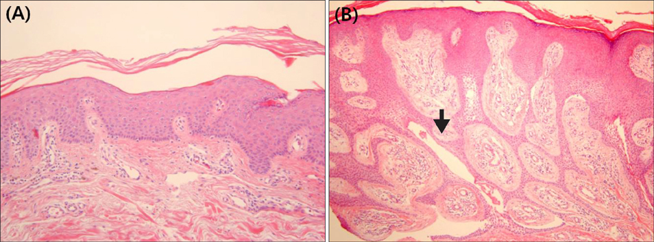
Fig. 2 Histopathologically, acanthosis, hyperkeratosis with exfoliation and perivascular inflammatory infiltration are observed in the superficial dermis (H&E, ×100). (B) Reticular, thin anastomosing strands of uniform cuboidal epithelial cells indicated in the arrow are growing into the dermis with epithelial cords embedded in a fibrovascular stroma (H&E, ×200).
Reference
-
1. Lui H, Stewart WD, English JC, Wood WS. Eccrine syringofibroadenomatosis: a clinical and histologic study and review of the literature. J Am Acad Dermatol. 1992; 26:805–813.
Article2. Cota C, Ferrara G, Amantea A, Donati P. Eccrine syringofibroadenoma and clear cell acanthoma: an association by chance? Am J Dermatopathol. 2011; 33:195–198.
Article3. Xu XL, Zhang W, Liu WD, Sun JF. A case of multiple eccrine syringofibroadenoma mimicking verruca vulgaris. J Dermatol. 2013; 40:665–666.
Article4. Starink TM. Eccrine syringofibroadenoma: multiple lesions representing a new cutaneous marker of the Schöpf syndrome, and solitary nonhereditary tumors. J Am Acad Dermatol. 1997; 36:569–576.
Article5. French LE. Reactive eccrine syringofibroadenoma: an emerging subtype. Dermatology. 1997; 195:309–310.
Article6. Utani A, Yabunami H, Kakuta T, Endo H, Shinkai H. Reactive eccrine syringofibroadenoma: an association with chronic foot ulcer in a patient with diabetes mellitus. J Am Acad Dermatol. 1999; 41:650–651.
Article7. Ichikawa E, Fujisawa Y, Tateishi Y, Imakado S, Otsuka F. Eccrine syringofibroadenoma in a patient with a burn scar ulcer. Br J Dermatol. 2000; 143:591–594.
Article8. Cho E, Lee JD, Cho SH. A case of reactive eccrine syringofibroadenoma. Ann Dermatol. 2011; 23:70–72.
Article9. Mehregan AH. Proliferation of sweat ducts in certain diseases of the skin. Am J Dermatopathol. 1981; 3:27–31.
Article10. Gambini C, Rongioletti F, Semino MT, Rebora A. Solitary eccrine syringofibroadenoma (or eccrine syringofibroadenomatous hyperplasia?) and diabetic polyneuropathy. Dermatology. 1996; 193:68–69.
Article11. Sueki H, Miller SJ, Dzubow LM, Murphy GF. Eccrine syringofibroadenoma (Mascaro): an ultrastructural study. J Cutan Pathol. 1992; 19:232–239.
Article12. Ohnishi T, Suzuki T, Watanabe S. Eccrine syringofibroadenoma. Report of a case and immunohistochemical study of keratin expression. Br J Dermatol. 1995; 133:449–454.
Article13. Takeda H, Mitsuhashi Y, Yoshikawa K, Katagata Y, Kondo S. Eccrine syringofibroadenoma: report of a case and analysis of cytokeratin expression. Dermatology. 1998; 196:242–245.
Article14. Saggini A, Mully T. Reactive eccrine syringofibroadenomatosis secondary to primary cutaneous amyloidosis: a novel association. J Cutan Pathol. 2014; 41:380–385.
Article15. Tey HL. Characterizing the nature of eccrine syringofibroadenoma: illustration with a case showing spontaneous involution. Clin Exp Dermatol. 2009; 34:e66–e68.
Article16. Bjarke T, Ternesten-Bratel A, Hedblad M, Rausing A. Carcinoma and eccrine syringofibroadenoma: a report of five cases. J Cutan Pathol. 2003; 30:382–392.
Article17. González-Serva A, Pró-Rísquez MA, Oliver M, Caruso MG. Syringofibrocarcinoma versus squamous cell carcinoma involving syringofibroadenoma: is there a malignant counterpart of Mascaro's syringofibroadenoma? Am J Dermatopathol. 1997; 19:58–65.
Article18. Lele SM, Gloster ES, Heilman ER, Chen PC, Chen CK, Anzil AP, et al. Eccrine syringofibroadenoma surrounding a squamous cell carcinoma: a case report. J Cutan Pathol. 1997; 24:193–196.
Article19. Mcniff J. Benign tumors with apocrine and eccrine differentiation. In : LeBoit PE, Weedon D, Sarason A, editors. World Health Organization Classification of Tumors. Pathology and genetics of skin tumors. IARC Press: Lyon;2006. p. 142.20. Trauner MA, Narurkar VA, Ruben BS. Eccrine syringofibroadenoma treated with a dual pulse width flashlamp pumped pulsed dye laser. Dermatol Surg. 1999; 25:418–420.
Article
