Ann Dermatol.
2017 Feb;29(1):95-99. 10.5021/ad.2017.29.1.95.
Multiple Skin Colored Nodules on both Legs in Patient with Positive QuantiFERON®-TB Gold Test
- Affiliations
-
- 1Department of Dermatology, Dankook University Medical College, Cheonan, Korea. misu2532@naver.com
- KMID: 2368036
- DOI: http://doi.org/10.5021/ad.2017.29.1.95
Abstract
- Nodular tuberculid (NT) was originally described by Jordaan et al. in 2000 in 4 patients from South Africa. It appeared as nodules on the legs; the pathologic changes were situated in the deep dermis and adjacent subcutaneous fat. A 34-year-old woman visited our hospital with subcutaneous skin-colored or slightly erythematous round to oval nodules. Skin biopsies revealed granulomatous inflammation at the dermo-subcutaneous junction with vasculitis. Chest X-ray, tuberculosus (TB)-polymerase chain reaction and TB culture of the skin specimen were normal. A QuantiFERON®-TB Gold test (QUIAGEN, Germany) was positive, which suggested a diagnosis of latent TB infection. The patient was treated with anti-TB medication and her condition has not recurred. Herein, we report a case of a patient with latent TB diagnosed by a positive QuantiFERON®-TB Gold test whose skin lesions had the clinical and histopathologic features of NT.
MeSH Terms
Figure
-
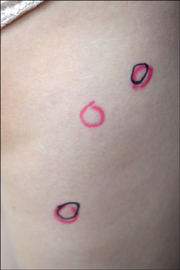
Fig. 1 Multiple tender skin-colored to slightly erythematous round to oval subcutaneous nodules.

Fig. 2 Granulomatous inflammation at the dermo-subcutaneous junction (H&E, ×40).
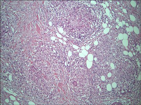
Fig. 3 Dense inflammatory cell infiltration around the vessels. Multinucleated giant cells, eosinophils, lymphocytes, foamy histiocytes, and neutrophils are evident (H&E, ×200).
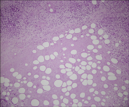
Fig. 4 Caseous necrosis in the superficial subcutaneous layer (H&E, ×100).
Reference
-
1. World Health Organization. Global tuberculosis report. World Health Organization;2014. Available from: http://apps.who.int/iris/bitstream/10665/137094/1/9789241564809_eng.pdf.2. World Health Organization. Global tuberculosis report 2013. World Health Organization;2013. Available from: http://apps.who.int/iris/bitstream/10665/91355/1/9789241564656_eng.pdf.3. Jordaan HF, Schneider JW, Abdulla EA. Nodular tuberculid: a report of four patients. Pediatr Dermatol. 2000; 17:183–188.
Article4. Asiniwasis R, Dutil MT, Walsh S. Molluscum-like papules as a presentation of early papulonecrotic tuberculid in association with nodular tuberculid in a male with asymptomatic active pulmonary tuberculosis. J Cutan Med Surg. 2015; 19:159–162.
Article5. Schneider JW, Jordaan HF, Geiger DH, Victor T, Van Helden PD, Rossouw DJ. Erythema induratum of Bazin. A clinicopathological study of 20 cases and detection of Mycobacterium tuberculosis DNA in skin lesions by polymerase chain reaction. Am J Dermatopathol. 1995; 17:350–356.
Article6. Goldsmith LA, Katz SI, Gilchrest BA, Paller AS, Leffell DJ, Wolff K. Fitzpatrick's dermatology in general medicine. 8th ed. New York: McGraw-Hill;2012. p. 2225–2240.7. Friedman PC, Husain S, Grossman ME. Nodular tuberculid in a patient with HIV. J Am Acad Dermatol. 2005; 53:2 Suppl 1. S154–S156.
Article8. Kumar B, Parsad D. Is ‘nodular tuberculid’ a distinct entity. Pediatr Dermatol. 2001; 18:164–167.
Article9. Lee JY, Shin SJ, Yoo CS, Kim CW, Kim SS. A case of atypical cutaneous tuberculid in an immunocompromised patient. Korean J Dermatol. 2013; 51:705–708.10. Lee JI, Park HJ, Lee JY, Cho BK. A case of lichen scrofulosorum developing after BCG vaccination. Korean J Dermatol. 2009; 47:309–312.11. Choi SJ, Won CH, Chang S, Lee MW, Choi JH, Moon KC. Lichen scrofulosorum occurring in association with tuberculous salpingitis. Korean J Dermatol. 2012; 50:358–361.12. Oh ST, Lee HJ, Youn DH, Kim JW. A case of papulonecrotic tuberculid combined with erythema induratum. Korean J Dermatol. 1999; 37:1703–1705.13. Yim JH, Koh BK, Kim JW. A case of papular tuberculid and BCGitis after BCG vaccination. Korean J Dermatol. 2005; 43:830–833.14. Han TY, Kim JY, Kwak HW, Choi JC, Shin JW, Kim JY, et al. A case of tuberculous lymphadenitis accompanying papulonecrotic tuberculid. Tuberc Respir Dis. 2007; 62:536–539.
Article15. Lee HJ, Park CJ, Yi JY, Kim TY, Kim CW. A case of papulonecrotic tuberculid combined with erythema nodosum. Ann Dermatol. 1995; 7:278–281.
Article16. Kim G, Jeong YI, Huh JW, Kim EJ, Joh OJ. Interferon-gamma release assay in a patient with tuberculosis verrucosa cutis. Ann Dermatol. 2015; 27:109–110.
Article17. Pai M, Riley LW, Colford JM Jr. Interferon-gamma assays in the immunodiagnosis of tuberculosis: a systematic review. Lancet Infect Dis. 2004; 4:761–776.
Article18. Angus J, Roberts C, Kulkarni K, Leach I, Murphy R. Usefulness of the QuantiFERON test in the confirmation of latent tuberculosis in association with erythema induratum. Br J Dermatol. 2007; 157:1293–1294.
Article
- Full Text Links
-
- Actions
-
Cited
- CITED
-
- Close
- Share
-
- Similar articles
-
- Comparison of Interferon-gamma Assays with the Tuberculin Skin Test in Children
- Application of the QuantiFERON(R)-TB Gold Test in Two Cases of Erythema Induratum of Bazin
- The Significance of Mantoux Test and Antitubercular Therapy in the Patients with Inflammatory Nodules on the Legs
- Tuberculin Skin Test and QuantiFERON-TB Gold Assay before and after Treatment for Latent Tuberculosis Infection among Health Care Workers in Local Tertiary Hospital
- The Diagnostic Value of Interferon-gamma Assay in Patients with Active Tuberculosis
