Ann Dermatol.
2017 Feb;29(1):69-73. 10.5021/ad.2017.29.1.69.
Digital Mucous Cyst: A Clinical-Surgical Study
- Affiliations
-
- 1Department of Dermatology, Wonkwang University School of Medicine, Iksan, Korea.
- 2Department of Dermatology, VHS Medical Center, Seoul, Korea. parkhjmd@medimail.co.kr
- KMID: 2368030
- DOI: http://doi.org/10.5021/ad.2017.29.1.69
Abstract
- BACKGROUND
It has been suggested digital mucous cysts (DMCs) are associated with osteoarthritis and osteophytes in the elderly, and usually have a communicating pedicle with the joint. Surgical excision is a standard therapy with a high cure rate.
OBJECTIVE
The purpose of this prospective study is to evaluate the features of DMCs via clinical, radiological and pathological examination and the efficacy of surgical excision of DMCs.
METHODS
Between 2010 and 2014, 24 Korean patients were treated with the resection of the cyst and the pedicle. Preoperative X-ray and ultrasonography were performed to detect the presence of the osteophyte and the connection to the joint space. Postoperative patients' satisfaction score was assessed by the visual analogue scale (0~10).
RESULTS
The osteophytes were found in 15.8%. In ultrasonographic findings, there were prominent flow signals between the cyst and the joint space in 13.6%. There were no serious postoperative complications, and recurrences were observed in 16.7%. Mean postoperative satisfaction score was 8.3.
CONCLUSION
It seems that preoperative X-ray for osteophytes and ultrasonographic study for connection are not always helpful for the treatment of DMCs, and that the surgical excision with a pedicle ligation and electrocoagulation is an effective treatment modality.
Keyword
MeSH Terms
Figure
-

Fig. 1 The surgical treatments of digital mucous cysts. (A) Before surgery, (B) dissected cyst, (C) pedicle (arrow), (D) after surgery.
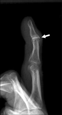
Fig. 2 The osteophytes (arrow) was found on X-rays (Case no. 21).

Fig. 3 On the ultrasonographic finding, there was a prominent flow signal between the cyst and the joint space (Case no. 4).
Cited by 1 articles
-
The Zitelli bilobed flap for soft tissue coverage after mucoid cyst resection: a retrospective cohort study
Sangho Oh, Jaein Chung, Daegu Son
Arch Hand Microsurg. 2024;29(2):90-95. doi: 10.12790/ahm.24.0014.
Reference
-
1. Miller PK, Roenigk RK, Amadio PC. Focal mucinosis (myxoid cyst). Surgical therapy. J Dermatol Surg Oncol. 1992; 18:716–719.
Article2. Salasche SJ. Myxoid cysts of the proximal nail fold: a surgical approach. J Dermatol Surg Oncol. 1984; 10:35–39.
Article3. Zook EG. Ganglions of the distal interphalangeal joint. In : Krull EA, Zook EG, Baran R, Haneke E, editors. Nail surgery: a text and atlas. New York: Lippincott Williams & Wilkins;2001. p. 209–212.4. De Berker DA, Lawrence CM. Treatment of myxoid cysts. Dermatol Surg. 2001; 27:296–299.
Article5. De Berker D, Lawrence C. Ganglion of the distal interphalangeal joint (myxoid cyst): therapy by identification and repair of the leak of joint fluid. Arch Dermatol. 2001; 137:607–610.6. Epstein E. A simple technique for managing digital mucous cysts. Arch Dermatol. 1979; 115:1315–1316.
Article7. Minami S, Nakagawa N, Ito T, Sadanobu N, Lin Y, Natsuaki M, et al. A simple and effective technique for the cryotherapy of digital mucous cysts. Dermatol Surg. 2007; 33:1280–1282.
Article8. Huerter CJ, Wheeland RG, Bailin PL, Ratz JL. Treatment of digital myxoid cysts with carbon dioxide laser vaporization. J Dermatol Surg Oncol. 1987; 13:723–727.
Article9. Kim JH, Park JH, Jee H, Oh SH. Successful treatment of recurrent digital mucoid cysts using a 1,444-nm neodymium-doped yttrium aluminum garnet laser. Dermatol Surg. 2011; 37:1528–1530.
Article10. Hur J, Kim YS, Yeo KY, Kim JS, Yu HJ. A case of herpetiform appearance of digital mucous cysts. Ann Dermatol. 2010; 22:194–195.
Article11. Lin YC, Wu YH, Scher RK. Nail changes and association of osteoarthritis in digital myxoid cyst. Dermatol Surg. 2008; 34:364–369.
Article12. Mani-Sundaram D. Surgical correction of mucous cysts of the nail unit. Dermatol Surg. 2001; 27:267–268.
Article13. Lee HJ, Kim PT, Jeon IH, Kyung HS, Ra IH, Kim TK. Osteophyte excision without cyst excision for a mucous cyst of the finger. J Hand Surg Eur Vol. 2014; 39:258–261.
Article14. Lawrence C. Skin excision and osteophyte removal is not required in the surgical treatment of digital myxoid cysts. Arch Dermatol. 2005; 141:1560–1564.
Article15. Brown RE, Zook EG, Russell RC, Kucan JO, Smoot EC. Fingernail deformities secondary to ganglions of the distal interphalangeal joint (mucous cysts). Plast Reconstr Surg. 1991; 87:718–725.
Article16. Sobanko JF, Dagum AB, Davis IC, Kriegel DA. Soft tissue tumors of the hand. 1. Benign. Dermatol Surg. 2007; 33:651–667.
Article17. Eke U, Ahmed I, Ilchyshyn A. Proximal nail fold flap dissection for digital myxoid cysts-a seven year experience. Dermatol Surg. 2014; 40:206–208.
Article
- Full Text Links
-
- Actions
-
Cited
- CITED
-
- Close
- Share
-
- Similar articles
-
- A Case of Digital Mucous Cyst Treated by Multiple Puncture Method
- Digital Mucous Cyst: Unusual Loeation and Characteristic Histopathologic Findings
- A Case of Digital Mucous Cyst Treated by Minocycline Sclerotherapy
- Three Cases of Digital Mucous Cyst Treated with Sodium Tetradecyl Sulfate Sclerotherapy
- Multiple Digital Mucous Cysts in a Farmer
