Usefulness of Dermoscopy in the Differential Diagnosis of Ruptured and Unruptured Epidermal Cysts
- Affiliations
-
- 1Department of Dermatology, Kosin University College of Medicine, Busan, Korea. ksderm77@unitel.co.kr
- 2Miul Dermatologic Clinic, Busan, Korea.
- KMID: 2368025
- DOI: http://doi.org/10.5021/ad.2017.29.1.33
Abstract
- BACKGROUND
An epidermal cyst is a common keratin-filled epithelial-lined cyst. The treatment of choice for epidermal cysts is surgical excision. If the cyst becomes ruptured, incision and drainage with oral antibiotic therapy or intralesional steroid injection are required.
OBJECTIVE
To analyze the dermoscopic features that can differentiate between ruptured and unruptured epidermal cysts.
METHODS
The clinical and dermoscopic features of the pathologically confirmed epidermal cysts of two subgroups of 38 patients, 20 with unruptured cysts and 18 with ruptured cysts, were reviewed.
RESULTS
With regard to the dermoscopic features, an ivory- white background color and punctum were commonly found in both groups (p>0.05). The unruptured-cyst group showed higher frequencies of pore sign (p<0.05), blue-white veil (p>0.05), no vascular structure, and arborizing telangiectasia (p<0.05), but the ruptured-cyst group usually had red lacunae (p>0.05) and peripheral linear branched vessels (with an erythematous rim) (p<0.05).
CONCLUSION
Dermoscopy is helpful in differentiating between ruptured and unruptured epidermal cysts.
Keyword
Figure
-
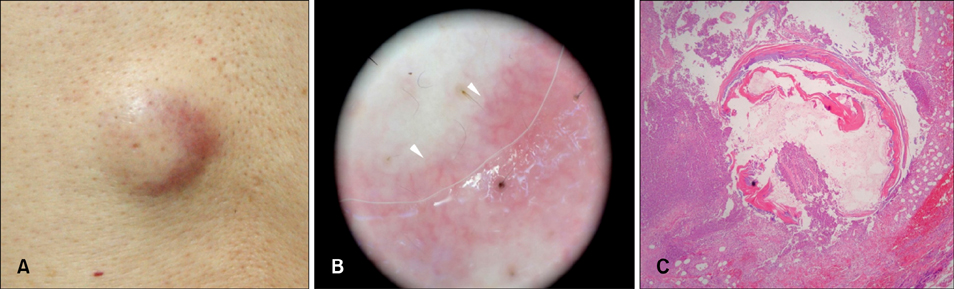
Fig. 1 Ruptured epidermal cyst. (A) Erythematous 2×2 cm subcutaneous nodule. (B) Dermoscopic view showing peripheral linear branched vessels with an erythematous rim (arrowheads) against an ivory-white background. (C) Microscopic view showing a cystic lesion with dense inflammation surrounding the ruptured cyst (patient 5 in the ruptured-cyst group).
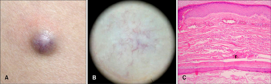
Fig. 2 Unruptured epidermal cyst. (A) Well-demarcated bluish 2×2 cm subcutaneous nodule. (B) Dermoscopic view showing a arborizing telangiectasia consisting of vessels with different diameter and several branches. (C) Microscopic view showing prominent dilated vessels with congestion of blood flow between the epidermis and cystic wall (patient 9 in the unruptured-cyst group).

Fig. 3 Schematic representation of dermoscopic pattern in patient with unruptured cyst and ruptured cyst.
Reference
-
1. Thomas VD, Snavely NR, Lee KK, Swanson NA. Benign epithelial tumors, hamartomas, and hyperplasias. Goldsmith LA, Katz SI, Gilchrest BA, Paller AS, Leffell DJ, Wolff K. Fitzpatrick's dermatology in general medicine. 8th ed. New York: McGraw Hill;2012. p. 1332–1334.2. Kirkham N. Tumors and cysts of the epidermis. Elder DE, Elenitsas R, Johnson BL, Murphy GF, Xu X. Lever's histopathology of the skin. 10th ed. Philadelphia: Lippincott Williams & Wilkins;2009. p. 800–802.3. Lee SY, Moon KC. A clinical and histopathologic study of epidermal cysts. Ann Dermatol. 1994; 6:157–161.
Article4. Chung J, Lee BJ, Ahn SK, Song DH, Lee WS, Kim SC. A clinical and histopathologic study of epidermal cysts. Korean J Dermatol. 1993; 31:517–522.5. Kim SK, Kwon H, Lee SY, Lee JS, Whang KU, Park YL, et al. A clinical and histopathological study of epidermal cysts in the province of Chungcheongnam-do. Korean J Dermatol. 2009; 47:516–523.6. McGavran MH, Binnington B. Keratinous cysts of the skin. Identification and differentiation of pilar cysts from epidermal cysts. Arch Dermatol. 1966; 94:499–508.
Article7. Cho HM, Kim SN. A clinical and histopathological study of 324 cases of epidermal cyst. Korean J Dermatol. 2007; 45:242–248.8. Lee HS, Joo KB, Song HT, Kim YS, Park DW, Park CK, et al. Relationship between sonographic and pathologic findings in epidermal inclusion cysts. J Clin Ultrasound. 2001; 29:374–383.
Article9. Jin W, Ryu KN, Kim GY, Kim HC, Lee JH, Park JS. Sonographic findings of ruptured epidermal inclusion cysts in superficial soft tissue: emphasis on shapes, pericystic changes, and pericystic vascularity. J Ultrasound Med. 2008; 27:171–176. quiz 177-178.
Article10. Benvenuto-Andrade C, Dusza SW, Agero AL, Scope A, Rajadhyaksha M, Halpern AC, et al. Differences between polarized light dermoscopy and immersion contact dermoscopy for the evaluation of skin lesions. Arch Dermatol. 2007; 143:329–338.
Article11. Pan Y, Gareau DS, Scope A, Rajadhyaksha M, Mullani NA, Marghoob AA. Polarized and nonpolarized dermoscopy: the explanation for the observed differences. Arch Dermatol. 2008; 144:828–829.12. Lallas A, Moscarella E, Argenziano G, Longo C, Apalla Z, Ferrara G, et al. Dermoscopy of uncommon skin tumours. Australas J Dermatol. 2014; 55:53–62.
Article13. Chandrasekaran V, Parkash S, Raghuveer CV. Epidermal cysts-a clinicopathological and biochemical study. Postgrad Med J. 1980; 56:823–827.14. Ghigliotti G, Cinotti E, Parodi A. Usefulness of dermoscopy for the diagnosis of epidermal cyst: the ‘pore’ sign. Clin Exp Dermatol. 2014; 39:649–650.
Article15. Park JS, Ko DK. A histopathologic study of epidermoid cysts in Korea: comparison between ruptured and unruptured epidermal cyst. Int J Clin Exp Pathol. 2013; 6:242–248.16. Pizzichetta MA, Canzonieri V, Soyer PH, Rubegni P, Talamini R, Massone C. Negative pigment network and shiny white streaks: a dermoscopic-pathological correlation study. Am J Dermatopathol. 2014; 36:433–438.17. Soyer HP, Argenziano G, Chimenti S, Ruocco V. Dermoscopy of pigmented skin lesions (Part XMLLink_XYZ). Eur J Dermatol. 2001; 11:483–498.
- Full Text Links
-
- Actions
-
Cited
- CITED
-
- Close
- Share
-
- Similar articles
-
- Usefulness of Ultrasonography in Differential Diagnosis between Ruptured and Unruptured Epidermal Cysts
- Usefulness of strain elastography in the differential diagnosis of ruptured epidermal cyst and superficial abscess
- A Ruptured Epidermal Inclusion Cyst in the Breast Presenting as a Recurrent Abscess
- Acquired Lymphangioma Circumscriptum of Vulva Mimicking Genital Wart: The Utility of Dermoscopy in Differential Diagnosis
- Angiographic Characteristics of the Intracranial Saccular Aneurysms to Predict the Rupture
