Restor Dent Endod.
2017 Feb;42(1):48-53. 10.5395/rde.2017.42.1.48.
Comparison of two different methods of detecting residual caries
- Affiliations
-
- 1Department of Restorative Dentistry, Hacettepe University, Sıhhıye, Ankara, Turkey. uzaykoc@gmail.com
- KMID: 2367321
- DOI: http://doi.org/10.5395/rde.2017.42.1.48
Abstract
OBJECTIVES
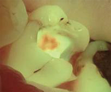
The aim of this study was to investigate the ability of the fluorescence-aided caries excavation (FACE) device to detect residual caries by comparing conventional methods in vivo.
MATERIALS AND METHODS
A total of 301 females and 202 males with carious teeth participated in this study. The cavity preparations were done by grade 4 (Group 1, 154 teeth), grade 5 (Group 2, 176 teeth), and postgraduate (Group 3, 173 teeth) students. After caries excavation using a handpiece and hand instruments, the presence of residual caries was evaluated by 2 investigators who were previously calibrated for visual-tactile assessment with and without magnifying glasses and trained in the use of a FACE device. The tooth number, cavity type, and presence or absence of residual caries were recorded. The data were analyzed using the Chi-square test, the Fisher's Exact test, or the McNemar test as appropriate. Kappa statistics was used for calibration. In all tests, the level of significance was set at p = 0.05.
RESULTS
Almost half of the cavities prepared were Class II (Class I, 20.9%; Class II, 48.9%; Class III, 20.1%; Class IV, 3.4%; Class V, 6.8%). Higher numbers of cavities left with caries were observed in Groups 1 and 2 than in Group 3 for all examination methods. Significant differences were found between visual inspection with or without magnifying glasses and inspection with a FACE device for all groups (p < 0.001). More residual caries were detected through inspection with a FACE device (46.5%) than through either visual inspection (31.8%) or inspection with a magnifying glass (37.6%).
CONCLUSIONS
Within the limitations of this study, the FACE device may be an effective method for the detection of residual caries.
Keyword
Figure
Reference
-
1. de Almeida Neves A, Coutinho E, Cardoso MV, Lambrechts P, Van Meerbeek B. Current concepts and techniques for caries excavation and adhesion to residual dentin. J Adhes Dent. 2011; 13:7–22.2. Ericson D, Kidd E, McComb D, Mjör I, Noack MJ. Minimally invasive dentistry-concepts and techniques in cariology. Oral Health Prev Dent. 2003; 1:59–72.3. Krause F, Braun A, Eberhard J, Jepsen S. Laser fluorescence measurements compared to electrical resistance of residual dentine in excavated cavities in vivo. Caries Res. 2007; 41:135–140.
Article4. Neves AA, Coutinho E, De Munck J, Lambrechts P, Van Meerbeek B. Does DIAGNOdent provide a reliable caries-removal endpoint? J Dent. 2011; 39:351–360.
Article5. Celiberti P, Francescut P, Lussi A. Performance of four dentine excavation methods in deciduous teeth. Caries Res. 2006; 40:117–123.
Article6. Banerjee A, Yasseri M, Munson M. A method for the detection and quantification of bacteria in human carious dentine using fluorescent in situ hybridisation. J Dent. 2002; 30:359–363.
Article7. Ismail AI. Visual and visuo-tactile detection of dental caries. J Dent Res. 2004; 83:Supplement C. C56–C66.
Article8. Nadanovsky P, Cohen Carneiro F, Souza de Mello F. Removal of caries using only hand instruments: a comparison of mechanical and chemo-mechanical methods. Caries Res. 2001; 35:384–389.
Article9. Neuhaus KW, Ellwood R, Lussi A, Pitts NB. Traditional lesion detection aids. Monogr Oral Sci. 2009; 21:42–51.
Article10. Lennon AM, Attin T, Martens S, Buchalla W. Fluorescence-aided caries excavation (FACE), caries detector, and conventional caries excavation in primary teeth. Pediatr Dent. 2009; 31:316–319.11. Coulthwaite L, Pretty IA, Smith PW, Higham SM, Verran J. The microbiological origin of fluorescence observed in plaque on dentures during QLF analysis. Caries Res. 2006; 40:112–116.
Article12. Koenig K, Schneckenburger H. Laser-induced autofluorescence for medical diagnosis. J Fluoresc. 1994; 4:17–40.
Article13. Lai G, Zhu L, Xu X, Kunzelmann KH. An in vitro comparison of fluorescence-aided caries excavation and conventional excavation by microhardness testing. Clin Oral Investig. 2014; 18:599–605.
Article14. Lai G, Kaisarly D, Xu X, Kunzelmann KH. MicroCT-based comparison between fluorescence-aided caries Detection of residual caries excavation and conventional excavation. Am J Dent. 2014; 27:12–16.15. Neves Ade A, Coutinho E, De Munck J, Van Meerbeek B. Caries-removal effectiveness and minimal-invasiveness potential of caries-excavation techniques: a micro-CT investigation. J Dent. 2011; 39:154–162.
Article16. Zhang X, Tu R, Yin W, Zhou X, Li X, Hu D. Micro-computerized tomography assessment of fluorescence aided caries excavation (FACE) technology: comparison with three other caries removal techniques. Aust Dent J. 2013; 58:461–467.
Article17. Unlu N, Ermis RB, Sener S, Kucukyilmaz E, Cetin AR. An in vitro comparison of different diagnostic methods in detection of residual dentinal caries. Int J Dent. 2010; 2010:864935.18. Meller C, Heyduck C, Tranaeus S, Splieth C. A new in vivo method for measuring caries activity using quantitative light-induced fluorescence. Caries Res. 2006; 40:90–96.
Article19. Ganter P, Al-Ahmad A, Wrbas KT, Hellwig E, Altenburger MJ. The use of computer-assisted FACE for minimal-invasive caries excavation. Clin Oral Investig. 2014; 18:745–751.
Article20. Lennon AM, Buchalla W, Switalski L, Stookey GK. Residual caries detection using visible fluorescence. Caries Res. 2002; 36:315–319.
Article21. Lennon AM, Attin T, Buchalla W. Quantity of remaining bacteria and cavity size after excavation with FACE, caries detector dye and conventional excavation in vitro. Oper Dent. 2007; 32:236–241.
Article22. Lennon AM, Buchalla W, Rassner B, Becker K, Attin T. Efficiency of 4 caries excavation methods compared. Oper Dent. 2006; 31:551–555.
Article23. Peskersoy C, Turkun M, Onal B. Comparative clinical evaluation of the efficacy of a new method for caries diagnosis and excavation. J Conserv Dent. 2015; 18:364–368.
Article24. Narula K, Kundabala M, Shetty N, Shenoy R. Evaluation of tooth preparations for Class II cavities using magnification loupes among dental interns and final year BDS students in preclinical laboratory. J Conserv Dent. 2015; 18:284–287.
Article25. Maggio MP, Villegas H, Blatz MB. The effect of magnification loupes on the performance of preclinical dental students. Quintessence Int. 2011; 42:45–55.26. Farook SA, Stokes RJ, Davis AK, Sneddon K, Collyer J. Use of dental loupes among dental trainers and trainees in the UK. J Investig Clin Dent. 2013; 4:120–123.
Article27. Zaugg B, Stassinakis A, Hotz P. Influence of magnification tools on the recognition of simulated preparation and filling errors. Schweiz Monatsschr Zahnmed. 2004; 114:890–896.
- Full Text Links
- Actions
-
Cited
- CITED
-
- Close
- Share
- Similar articles
-
- Comparison of Diagnostic Validity between Laser Fluorescence Devices in Proximal Caries
- Detecting of Proximal Caries in Primary Molars using Pen-type QLF Device
- Deep learning convolutional neural network algorithms for the early detection and diagnosis of dental caries on periapical radiographs: A systematic review
- Assessment of the Object Detection Ability of Interproximal Caries on Primary Teeth in Periapical Radiographs Using Deep Learning Algorithms
- Digital contrast subtraction radiography for proximal caries diagnosis


