Investig Magn Reson Imaging.
2016 Dec;20(4):250-253. 10.13104/imri.2016.20.4.250.
An Unusual Case of Japanese Encephalitis Involving Unilateral Deep Gray Matter and Temporal Lobe on Diffusion-Weighted MRI
- Affiliations
-
- 1Department of Radiology, Seoul Medical Center, Seoul, Korea. jnoon276@gmail.com
- KMID: 2366407
- DOI: http://doi.org/10.13104/imri.2016.20.4.250
Abstract
- Acute Japanese encephalitis (JE) is an endemic viral infectious disease in various parts of Far East and Southeast Asian countries including Korea. Bilateral thalami are the most common involving sites in JE. Other areas including the basal ganglia, substantia nigra, red nucleus, pons, cerebral cortex and cerebellum may be also involved. We report an extremely unusual brain diffusion-weighted MR imaging (DWI) findings in a 53-year-old man with serologically proven JE involving unilateral deep gray matter and temporal lobe, which shows multifocal high signal intensities in left thalamus, left substantia nigra, left caudate nucleus and left medial temporal cortex on T2-weighted image and DWI with iso-intensity on apparent diffusion coefficient (ADC) map.
Keyword
MeSH Terms
Figure
-
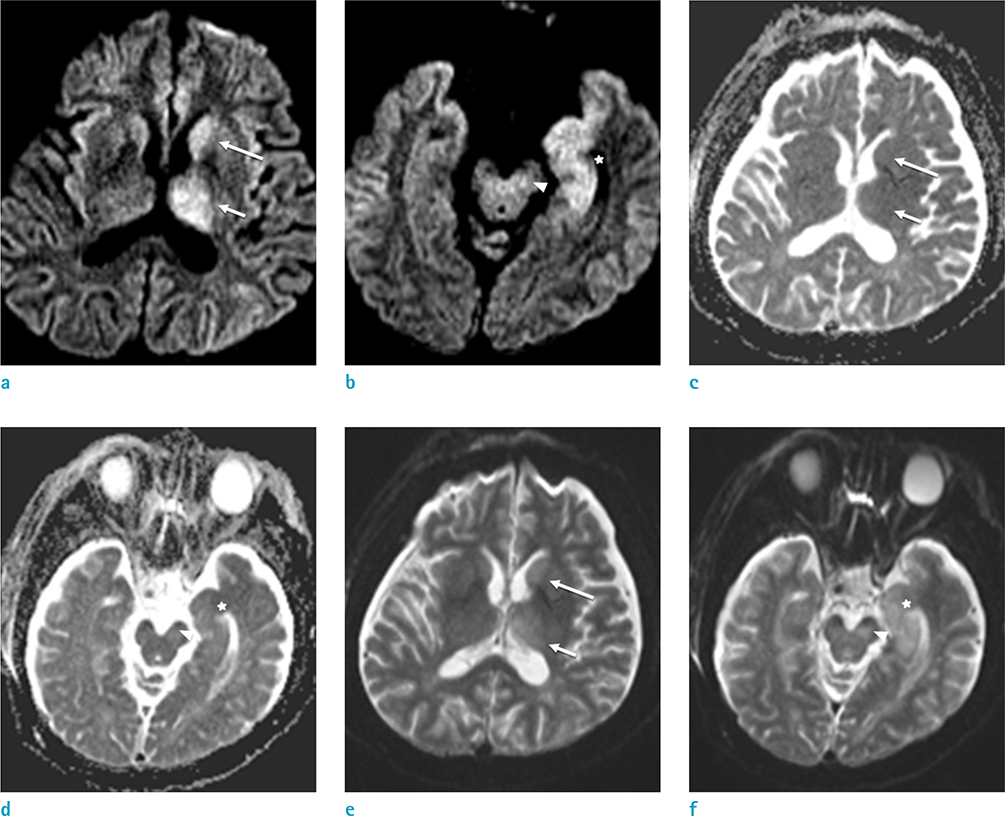
Fig. 1 A 53-year-old man with JE. Diffusion-weighted imaging (DWI) (b = 1000 s/mm2) (a, b), apparent diffusion coefficient (ADC) map (c, d) and T2-weighted image (DWI b = 0 s/mm2) (e, f) obtained on day 2 after onset of altered mentality show multiple areas of high intensity on DWI (b = 1000 s/mm2) (a, b) and isointensity on ADC map (c, d) in the left thalamus (short arrow), left caudate nucleus (long arrow), left subtantia nigra (arrowhead) and left medial temporal lobe (asterisk). On T2-weighted image (DWI b = 0 s/mm2, e, f), the lesions show subtle high intensity.
Reference
-
1. Basumatary LJ, Raja D, Bhuyan D, Das M, Goswami M, Kayal AK. Clinical and radiological spectrum of Japanese encephalitis. J Neurol Sci. 2013; 325:15–21.2. Kalita J, Misra UK. Comparison of CT scan and MRI findings in the diagnosis of Japanese encephalitis. J Neurol Sci. 2000; 174:3–8.3. Kumar S, Misra UK, Kalita J, Salwani V, Gupta RK, Gujral R. MRI in Japanese encephalitis. Neuroradiology. 1997; 39:180–184.4. Yakushiji Y, Kurohara K, Tanaka A, Kuroda Y, Uchino A. A case of Japanese encephalitis presenting with unilateral lesions in diffusion-weighted MRI. Rinsho Shinkeigaku. 2001; 41:602–605.5. Solomon T. Viral encephalitis in Southeast Asia. Neurol Infect Epidemiol. 1997; 2:191–199.6. Handique SK, Das RR, Barman K, et al. Temporal lobe involvement in Japanese encephalitis: problems in differential diagnosis. AJNR Am J Neuroradiol. 2006; 27:1027–1031.7. Sawlani V. Diffusion-weighted imaging and apparent diffusion coefficient evaluation of herpes simplex encephalitis and Japanese encephalitis. J Neurol Sci. 2009; 287:221–226.8. Hegde AN, Mohan S, Lath N, Lim CC. Differential diagnosis for bilateral abnormalities of the basal ganglia and thalamus. Radiographics. 2011; 31:5–30.9. Meissner B, Kallenberg K, Sanchez-Juan P, et al. Isolated cortical signal increase on MR imaging as a frequent lesion pattern in sporadic Creutzfeldt-Jakob disease. AJNR Am J Neuroradiol. 2008; 29:1519–1524.10. Zeidler M, Sellar RJ, Collie DA, et al. The pulvinar sign on magnetic resonance imaging in variant Creutzfeldt-Jakob disease. Lancet. 2000; 355:1412–1418.
- Full Text Links
-
- Actions
-
Cited
- CITED
-
- Close
- Share
-
- Similar articles
-
- Diffusion Tensor Imaging of Heterotopia: Changes of Fractional Anisotropy during Radial Migration of Neurons
- Japanese Encephalitis Associated with Flaccid Quadriplegia
- Japanese Encephalitis Presenting with Unilateral Medial Temporal Lobe Lesion
- Diffusion-Weighted Imaging of Brain Injury Due to Neonatal Hypoglycemia: A Case Report
- Epstein-Barr Virus Encephalitis Involving the Bilateral Corticospinal Tract: Wine Glass Sign
