Malignant Transformation of Craniopharyngioma without Radiation Therapy: Case Report and Review of the Literature
- Affiliations
-
- 1Department of Neurosurgery, Gil Medical Center, Gachon University, Incheon, Korea. gtyee@gilhospital.com
- 2Department of Pathology, Gil Medical Center, Gachon University, Incheon, Korea.
- KMID: 2365596
- DOI: http://doi.org/10.3340/jkns.2015.0707.022
Abstract
- Craniopharyngiomas exhibiting histologic malignancy are extremely rare. Herein, we report the case of a 26-year-old male patient who underwent suprasellar mass excision via an interhemispheric transcallosal approach. Histopathological examination indicated that the craniopharyngioma was of the adamantinomatous subtype. The patient received postoperative medical treatment for endocrine dysfunction and diabetes mellitus without radiation treatment. Two years after the operation, he presented with progressive visual disturbance and altered mentality. Magnetic resonance imaging revealed a huge mass in the suprasellar cistern and third ventricle. He underwent a second operation via the same approach. The histopathological examination showed an adamantinomatous craniopharyngioma with sheets of solid proliferation in a spindled pattern, indicating malignant transformation. Malignant transformation of craniopharyngioma in the absence of radiation therapy has been reported in only five cases, including this one. We present a case of malignant transformation of craniopharyngioma with a brief review of relevant literature.
MeSH Terms
Figure
-

Fig. 1 Brain magnetic resonance imaging before the first surgery, showing a suprasellar mass lesion with enhanced solid and multiseptated cystic components. A : Axial. B : Coronal. C : Sagittal.
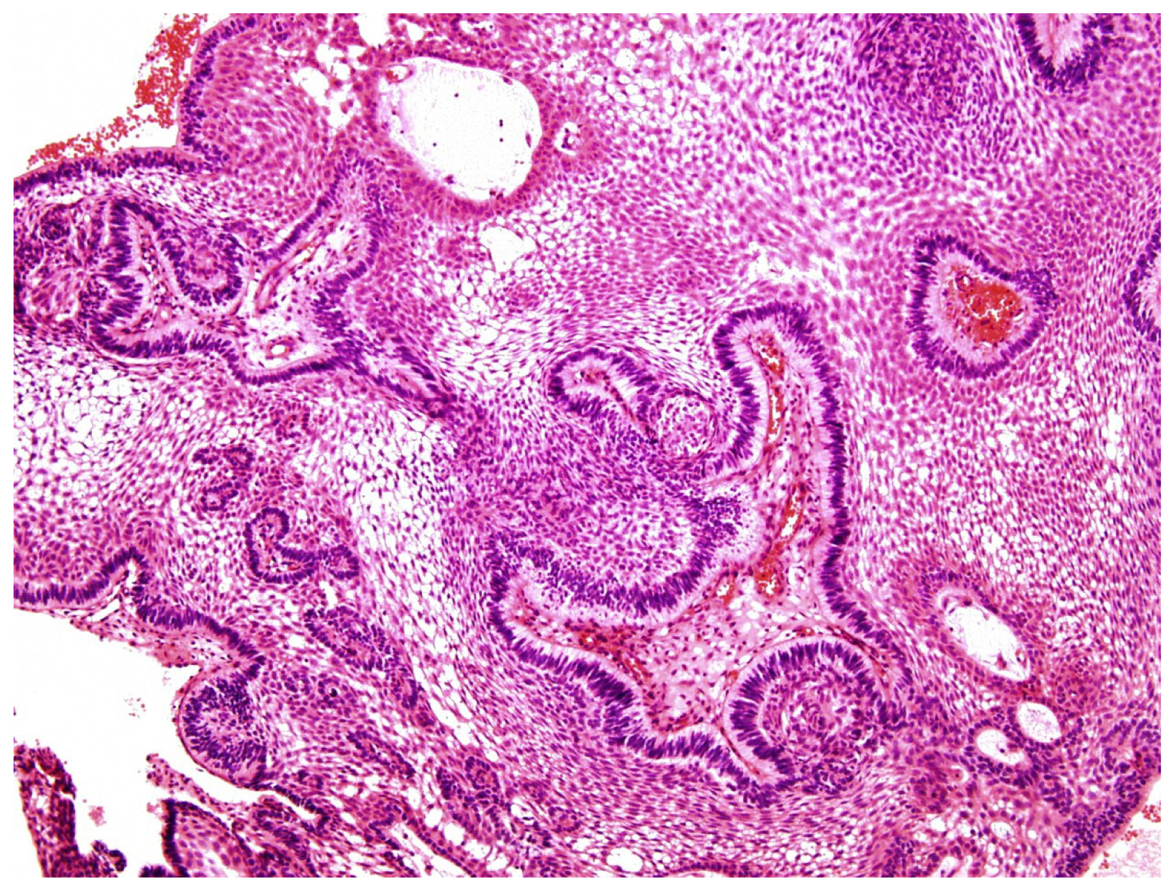
Fig. 2 The first biopsy showing a typical adamantinomatous craniopharyngioma.
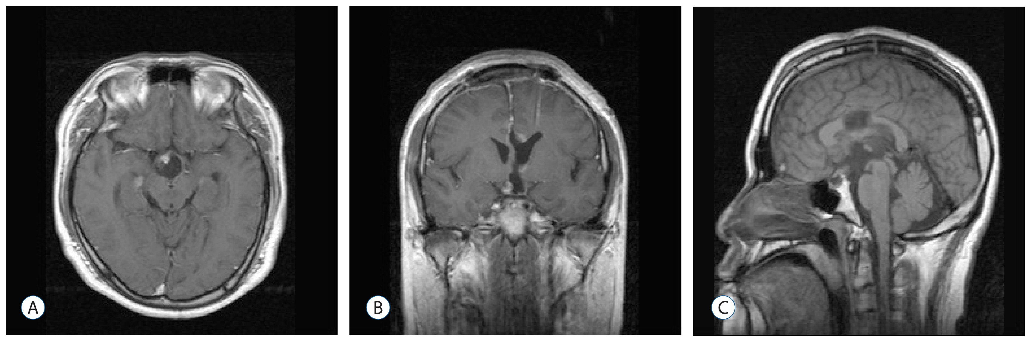
Fig. 3 Post-operative magnetic resonance imaging scan obtained 6 months after the first surgery, showing a small residual enhancing mass along the right anterior margin of the floor of the third ventricle. A : Axial. B : Coronal. C : Sagittal.
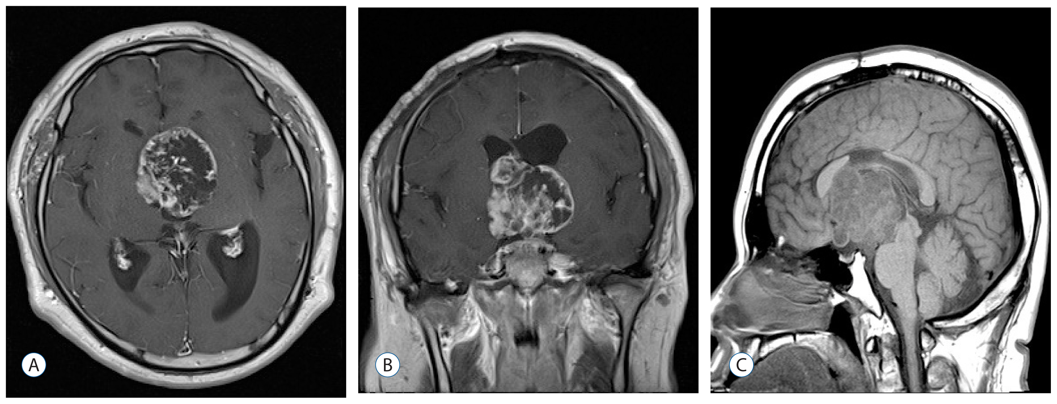
Fig. 4 Follow-up brain magnetic resonance imaging 2 years after the first surgery, showing recurrence of multiseptated huge cystic mass in the suprasellar, third, and lateral ventricles. A : Axial. B : Coronal. C : Sagittal.

Fig. 5 Histopathologic findings of a specimen obtained during the second surgery. Typical adamantinomatous craniopharyngioma that is highly cellular and presents with a spindled pattern (A, B; hematoxylin-eosin). The immunological tests showing overexpression of p53 (C) and Ki67 (D), and positive results for pancytokeratin (E) and vimentin (F).
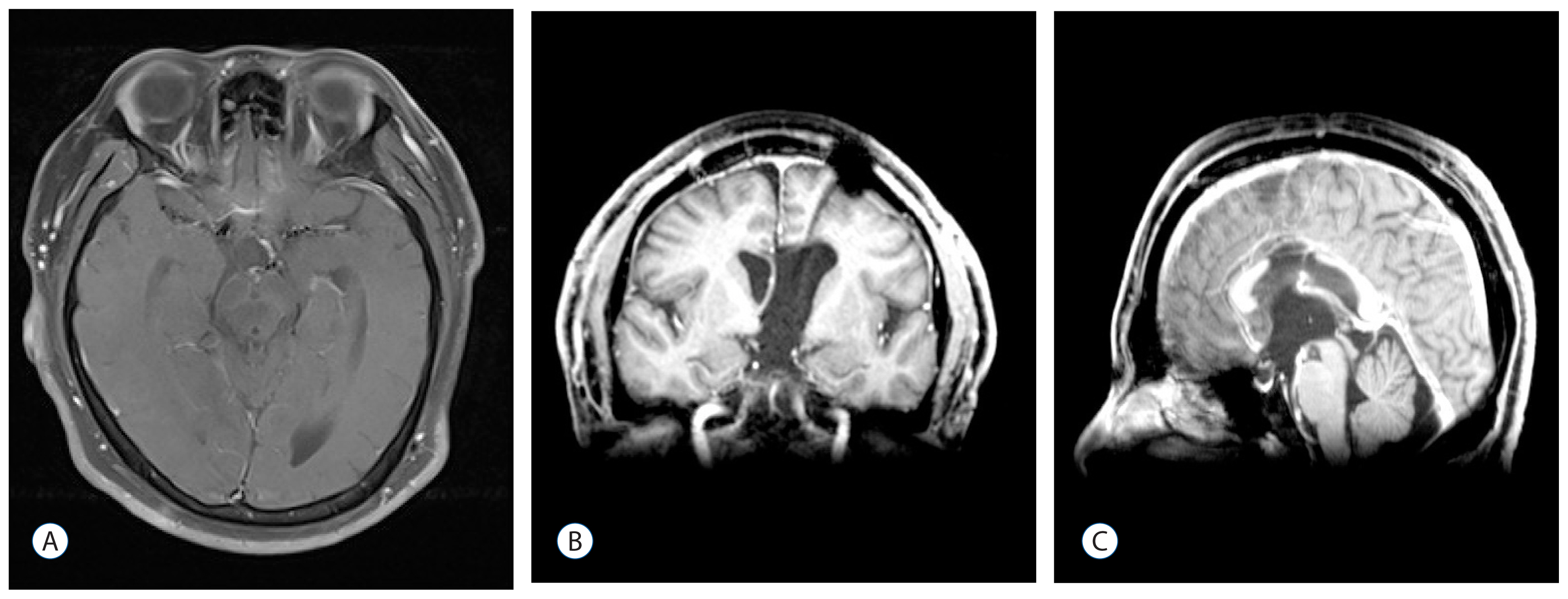
Fig. 6 Brain magnetic resonance imaging after the second surgery showing total resection of the recurrent mass. A : Axial. B : Coronal. C : Sagittal.
Reference
-
References
1. Akachi K, Takahashi H, Ishijima B, Nakamura Y, Oda M, Takizawa T, et al. Malignant changes in a craniopharyngioma. No Shinkei Geka. 15:843–848. 1987.2. Boongird A, Laothamatas J, Larbcharoensub N, Phudhichareonrat S. Malignant craniopharyngioma; case report and review of the literature. Neuropathology. 29:591–596. 2009.
Article3. Bunin GR, Surawicz TS, Witman PA, Preston-Martin S, Davis F, Bruner JM. The descriptive epidemiology of craniopharyngioma. J Neurosurg. 89:547–551. 1998.
Article4. Gao S, Shi X, Wang Y, Qian H, Liu C. Malignant transformation of craniopharyngioma: case report and review of the literature. J Neurooncol. 103:719–725. 2011.
Article5. Jaggon J, Abrikian S, Gibson T, Johnson P, Liburd J. Malignant craniopharyngioma: a case report and comprehensive review. Internet J Pathol. 11:1–4. 2009.6. Kohn HI, Fry RJ. Radiation carcinogenesis. New Engl J Med. 310:504–511. 1984.
Article7. Louis DN, Ohgaki H, Wiestler OD, Cavenee WK, Burger PC, Jouvet A, et al. The 2007 WHO classification of tumours of the central nervous system. Acta Neuropathol. 114:97–109. 2007.
Article8. Mark RJ, Lutge WR, Shimizu KT, Tran LM, Selch MT, Parker RG. Craniopharyngioma: treatment in the CT and MR imaging era. Radiology. 197:195–198. 1995.
Article9. Rodriguez FJ, Scheithauer BW, Tsunoda S, Kovacs K, Vidal S, Piepgras DG. The spectrum of malignancy in craniopharyngioma. Am J Surg Pathol. 31:1020–1028. 2007.
Article10. Salyer D, Carter D. Squamous carcinoma arising in the pituitary gland. Cancer. 31:713–718. 1973.
Article11. Wang W, Chen XD, Bai HM, Liao QL, Dai XJ, Peng DY, et al. Malignant transformation of craniopharyngioma with detailed follow-up. Neuropathology. 35:50–55. 2015.
Article12. Yue Y, Da JP. Malignant transformation of craniopharyngioma: a case report. Zhonghua Bing Li Xue Za Zhi. 35:439. 2006.
- Full Text Links
-
- Actions
-
Cited
- CITED
-
- Close
- Share
-
- Similar articles
-
- A Case of Papillary Transitional Cell Carcinoma Arising from the Benign Cystic Teratoma of Ovary
- A Case Report of Craniopharyngioma
- Postoperative Intratumoral Injection of Bleomycin for Cystic Craniopharyngioma
- Intracranial Anaplastic Astrocytoma after Radiotherapy for Craniopharyngioma
- Moyamoya Syndrome Precipitated by Cranial Irradiation for Craniopharyngioma in Children