Skull Metastasis of Gastric Gastrointestinal Stromal Tumor Successfully Managed by Surgery
- Affiliations
-
- 1Division of Hematology-Oncology, Department of Internal Medicine, Gil Medical Center, Gachon University College of Medicine, Incheon, Korea. dbs@gilhospital.com
- 2Department of Pathology, Gil Medical Center, Gachon University College of Medicine, Incheon, Korea.
- 3Department of Neurosurgery, Gil Medical Center, Gachon University College of Medicine, Incheon, Korea.
- KMID: 2365593
- DOI: http://doi.org/10.3340/jkns.2014.0506.007
Abstract
- Gastrointestinal stromal tumors (GISTs) are rare, but are the most common mesenchymal neoplasm of the gastrointestinal tract. The most common sites of metastasis are liver and peritoneum, while bone metastasis is rare. We report on a patient with skull metastasis after seven years of treatment with imatinib for metastatic GIST. She underwent metastasectomy consisting of craniectomy with excision of the mass, and cranioplasty and continued treatment with imatinib and sunitinib, without evidence of cranial recurrence. She died of pneumonia sepsis one year after metastasectomy. Skull metastasis of GIST is a very rare presentation, and an aggressive multidisciplinary approach should be considered whenever possible.
Keyword
MeSH Terms
Figure
-
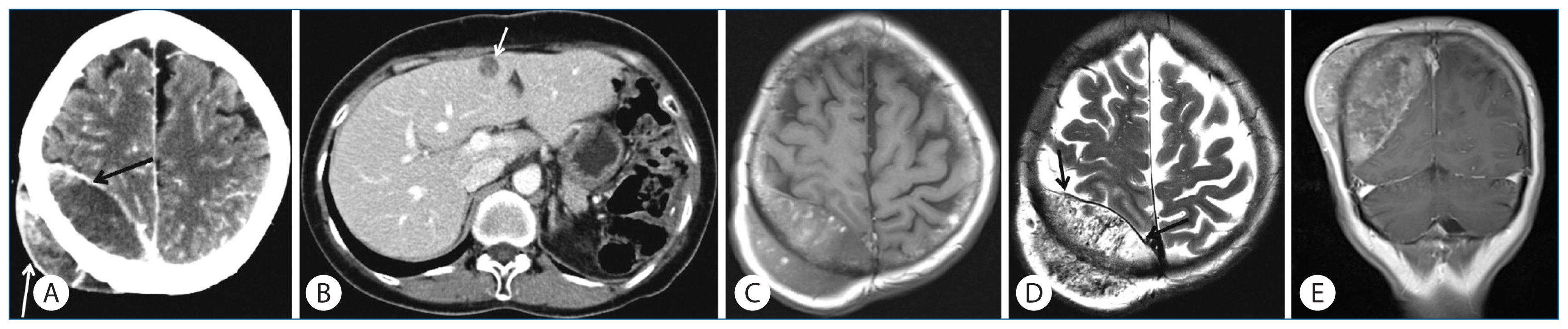
Fig. 1 Imaging findings of skull metastasis and hepatic metastasis in a patient with malignant gastrointestinal stromal tumor. A: Post-contrast brain CT image reveals a 7.7 cm sized, well-defined mass (arrows) centered on the diploic space of the calvaria. B: Abdomen CT image reveals a hypodense single metastasis (arrow) in left medial segment of the liver. C and D: Axial T1- (C) and T2-weighted (D) MR images show heterogeneous signal intensity of the skull metastasis. The dura (arrows) is intact and the underlying cortex appears compressed. Note the multiple signal voids on T2-weighted image. E: After gadolinium enhancement, the mass demonstrates heterogeneous enhancement.

Fig. 2 Pathology of a resected skull tumor. A: Metastatic gastrointestinal stromal tumor in the skull. The tumor consists of atypical spindle cells with high cellularity and infiltrative growth pattern with destruction of normal bone tissue (H&E, ×100). B: High power view of the tumor. Mitotic figures (arrows) are frequently noted (H&E, ×400). C: Tumor cells are positive for c-kit (c-kit immunostain, ×400).
Cited by 1 articles
-
Revisiting the Role of Surgical Resection for Brain Metastasis
Joonho Byun, Jong Hyun Kim
Brain Tumor Res Treat. 2023;11(1):1-7. doi: 10.14791/btrt.2022.0028.
Reference
-
References
1. Abuzakhm SM, Acre-Lara CE, Zhao W, Hitchcock C, Mohamed N, Arbogast D, et al. Unusual metastases of gastrointestinal stromal tumor and genotypic correlates: case report and review of the literature. J Gastrointest Oncol. 2:45–49. 2011.2. Akiyama K, Numaga J, Kagaya F, Takazawa Y, Suzuki S, Koseki N, et al. Case of optic nerve involvement in metastasis of a gastrointestinal stromal tumor. Jpn J Ophthalmol. 48:166–168. 2004.
Article3. Baeg MK, Bae SH, Lee KH, Kim J, Park IS, Jin JY. Diplopia as a presenting symptom in a gastric gastrointestinal stromal tumor. Jpn J Clin Oncol. 41:265–268. 2011.
Article4. Barrière J, Thariat J, Vandenbos F, Bondiau PY, Peyrottes I, Peyrade F. Diplopia as the first symptom of an aggressive metastatic rectal stromal tumor. Onkologie. 32:345–347. 2009.
Article5. Bertulli R, Fumagalli E, Coco P, Messina A, Morosi C, Dileo P, et al. Unusual metastatic sites in gastrointestinal stromal tumor (GIST). J Clin Oncol (Meeting Abstracts). 27:10566. 2009.
Article6. Di Scioscio V, Greco L, Pallotti MC, Pantaleo MA, Maleddu A, Nannini M, et al. Three cases of bone metastases in patients with gastrointestinal stromal tumors. Rare Tumors. 3:e17. 2011.
Article7. Eisenberg BL, Pipas JM. Gastrointestinal stromal tumor--background, pathology, treatment. Hematol Oncol Clin North Am. 26:1239–1259. 2012.
Article8. Feki J, Bouzguenda R, Ayedi L, Bradi M, Boudawara T, Daoud J, et al. Bone metastases from gastrointestinal stromal tumor: a case report. Case Rep Oncol Med. 2012:509845. 2012.
Article9. Gil-Arnaiz I, Martínez-Trufero J, Pazo-Cid RA, Felipo F, Lecumberri MJ, Calderero V. Skull metastasis from rectal gastrointestinal stromal tumours. Clin Transl Oncol. 11:625–627. 2009.
Article10. Jati A, Tatlı S, Morgan JA, Glickman JN, Demetri GD, Van den Abbele A, et al. Imaging features of bone metastases in patients with gastrointestinal stromal tumors. Diagn Interv Radiol. 18:391–396. 2012.11. Li LF, Tse YH, Ho SL, Yan KW, Lui WM. Duodenal GIST metastasized to skull and orbit managed by surgery: a case report. Asian J Surg. 34:181–184. 2011.
Article12. Raut CP, Posner M, Desai J, Morgan JA, George S, Zahrieh D, et al. Surgical management of advanced gastrointestinal stromal tumors after treatment with targeted systemic therapy using kinase inhibitors. J Clin Oncol. 24:2325–2331. 2006.
Article13. van der Zwan SM, DeMatteo RP. Gastrointestinal stromal tumor: 5 years later. Cancer. 104:1781–1788. 2005.
Article14. von Mehren M, Heinrich MC, Joensuu H, Blanke CD, Wehrle E, Demetri GD. Follow-up results after 9 years (yrs) of the ongoing, phase II B2222 trial of imatinib mesylate (IM) in patients (pts) with metastatic or unresectable KIT+ gastrointestinal stromal tumors (GIST). J Clin Oncol. 29(Suppl):Abstract 10016. 2011.
Article15. Wong CS, Chu YC. Intra-cranial metastasis of gastrointestinal stromal tumor. Chin Med J (Engl). 124:3595–3597. 2011.
- Full Text Links
-
- Actions
-
Cited
- CITED
-
- Close
- Share
-
- Similar articles
-
- A Ruptured Metastatic Hepatic Gastrointestinal Stromal Tumor Treated by Angiographic Embolization
- Gastrointestinal Stromal Tumor with Extensive Lymphatic Metastasis: A Case Report
- Extragastrointestinal Stromal Tumor Mimicking Gastric Subepithelial Tumor
- Systemic Treatment of the Gastrointestinal Stromal Tumor (GIST)
- A Case of Epithelioid Type Gastric Gastrointestinal Stromal Tumor with Gastrointestinal Bleeding
