Rapidly Growing Right Ventricular Outflow Tract Mass in Patient with Sarcomatoid Renal Cell Carcinoma
- Affiliations
-
- 1Department of Internal Medicine, Cardiovascular Center, Pusan National University Yangsan Hospital, Pusan National University School of Medicine, Yangsan, Korea. nadroj@chol.com
- 2Department of Pathology, Pusan National University Yangsan Hospital, Yangsan, Korea.
- KMID: 2364652
- DOI: http://doi.org/10.4250/jcu.2016.24.4.329
Abstract
- Cardiac metastasis from renal cell carcinoma (RCC) without inferior vena cava (IVC) involvements is extremely rare with few reported cases. Sarcomatoid RCC with rhabdoid feature is a rare pathologic type of RCC having aggressive behavior due to great metastatic potential. Here, we report a case of rapidly growing cardiac metastasis of RCC which brought on right ventricular outflow tract (RVOT) obstruction without IVC and right atrial involvement in a 61-year-old woman. Cardiac arrest occurred during radical nephrectomy and echocardiography revealed mass nearly obstructing the RVOT which was not recognized by preoperative echocardiography 1 month ago. Postoperative immunohistochemical evaluation of renal mass revealed sarcomatoid RCC with rhabdoid feature.
Keyword
MeSH Terms
Figure
-

Fig. 1 Abdominal computed tomography (CT) revealed 9-cm sized left renal mass (arrow) (A). Chest CT showed scanty amount of pericardial effusion (arrowheads) (B).
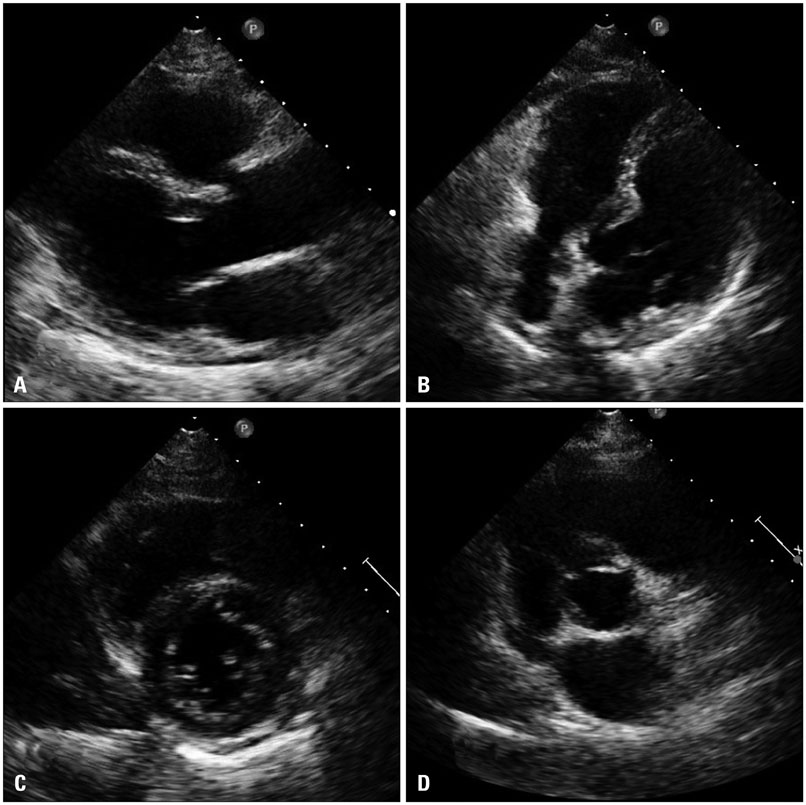
Fig. 2 Parasternal long axis view on transthoracic echocardiography showed normal left ventricle (LV) size and no evidence of right ventricular outflow tract mass (A). Modified four chamber view also showed normal LV and RV size with no evidence of intracardiac mass (B). In parasternal short axis view, no gross abnormality was observed (C and D). RV: right ventricle.
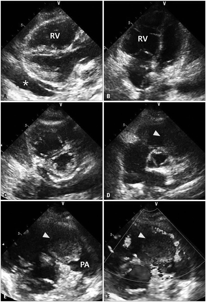
Fig. 3 Parasternal long axis view (A) and modified 4 chamber view (B) on transthoracic echocardiography revealed markedly dilated RV with moderate amount of pericardial effusion (asterisk). Parasternal short axis view showed D-shaped left ventricle (C). Parasternal short axis view of aortic valve level. In this view, right ventricular outflow tract (RVOT) mass (arrowhead) was hardly seen (D). Parasternal short axis view of RVOT level demonstrated 5.5 × 3 cm sized mass (arrowhead) nearly obstructing the RVOT (E). In this view, obstruction of blood flow by this RVOT mass (arrowhead) was well visualized under color Doppler image (F). RV: right ventricle, PA: pulmonary artery.

Fig. 4 The tumor showed an ill-defined, whitish, infiltrating mass with necrosis.
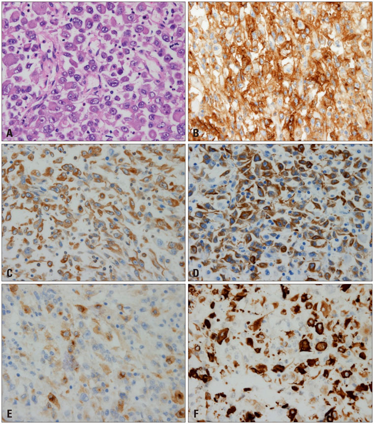
Fig. 5 The tumor composed of epithelioid tumor cells with rhabdoid feature. A: Hematoxylin and eosin staining (× 400). B: CD10 staining (× 400). C: Vimentin staining (× 400). D: Pan-cytokeratin staining (× 400). E: EMA staining (× 400). F: Desmin staining (× 400).
Reference
-
1. Zustovich F, Gottardo F, De Zorzi L, Cecchetto A, Dal Bianco M, Mauro E, Cartei G. Cardiac metastasis from renal cell carcinoma without inferior vena involvement: a review of the literature based on a case report. Two different patterns of spread? Int J Clin Oncol. 2008; 13:271–274.2. Ro JY, Ayala AG, Sella A, Samuels ML, Swanson DA. Sarcomatoid renal cell carcinoma: clinicopathologic. A study of 42 cases. Cancer. 1987; 59:516–526.3. Roberts WC. Primary and secondary neoplasms of the heart. Am J Cardiol. 1997; 80:671–682.4. Butz T, Schmidt HK, Fassbender D, Esdorn H, Wiemer M, Horstkotte D, Faber L. Echo-guided percutaneous coil embolization of a symptomatic massive metastasis of a renal cell carcinoma in the right ventricular outflow tract. Eur J Echocardiogr. 2008; 9:725–727.5. Shannon B, Stan Wisniewski Z, Bentel J, Cohen RJ. Adult rhabdoid renal cell carcinoma. Arch Pathol Lab Med. 2002; 126:1506–1510.6. Ding GT, Hwang JS, Tan PH. Sarcomatoid renal cell carcinoma metastatic to the breast: report of a case with diagnosis on fine needle aspiration cytology. Acta Cytol. 2007; 51:451–455.7. Zhao WP, Yu YL, Chen ZQ, Huang XF, Zhang ZG. Colon metastasis of chromophobe renal cell carcinoma with sarcomatoid change. Chin Med J (Engl). 2012; 125:2352–3354.8. Gammon BL, Gleason BC, Thomas AB, Cibull TL. Sarcomatoid renal cell carcinoma presenting in the oropharynx. J Cutan Pathol. 2010; 37:1255–1258.
- Full Text Links
-
- Actions
-
Cited
- CITED
-
- Close
- Share
-
- Similar articles
-
- A Case of Sarcomatoid Renal Cell Carcinoma with Multiple Renal Stones and Pyonephrosis
- Sarcomatoid Renal Cell Carcinoma; Special Reference to its Distinction from Carcinosarcoma
- Left Ventricular Metastasis From Renal Cell Carcinoma Causing Left Ventricular Outflow Tract Obstruction
- A Case of Vulvar Carcinoma: Squamous Cell Carcinoma with Sarcomatoid Features
- Sarcomatoid Renal Cell Carcinoma
