Proliferative periostitis of the mandibular ramus and condyle: a case report
- Affiliations
-
- 1Department of Oral and Maxillofacial Surgery, College of Dentistry, Gangneung-Wonju National University, Gangneung, Korea.
- 2Department of Oral and Maxillofacial Surgery, Jeju National University Hospital, Jeju National University School of Medicine, Jeju, Korea. 2460song@naver.com
- KMID: 2364017
- DOI: http://doi.org/10.5125/jkaoms.2015.41.4.198
Abstract
- Proliferative periostitis is a rare form of osteomyelitis that is characterized by new bone formation with periosteal reaction common causes of proliferative periostitis are dental caries, periodontitis, cysts, and trauma. While proliferative periostitis typically presents as a localized lesion, in this study, we describe an extensive form of proliferative periostitis involving the whole mandibular ramus and condyle. Because the radiographic findings were similar to osteogenic sarcoma, an accurate differential diagnosis was important for proper treatment.
Keyword
MeSH Terms
Figure
-
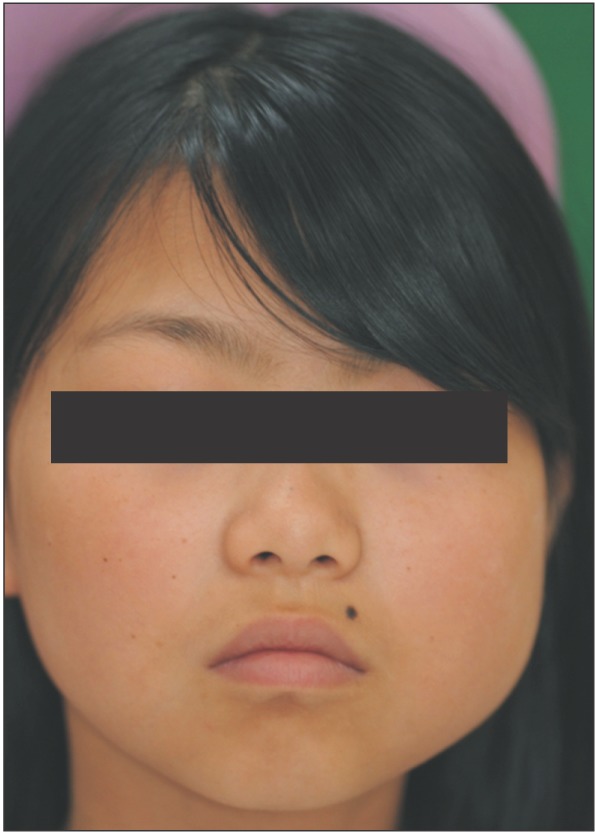
Fig. 1 Clinical photograph shows the swelling of the left mandibular ramus and angle.
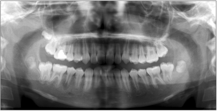
Fig. 2 Panoramic radiographs show the radiolucent appearance around the unerupted third molar on the left mandible.
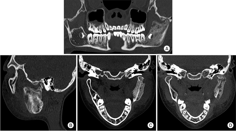
Fig. 3 Computed tomography image. Osteolytic and sclerotic bone lesion. A. The ramus and condyle. B. Mandibular angle. C. Periosteal reaction in the left mandibular ramus and condyle. D. Osteolytic change inside the newly formed bone.
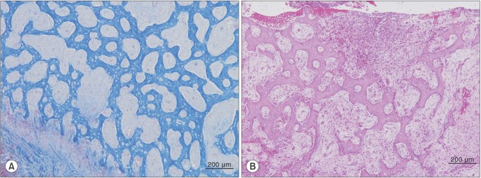
Fig. 4 Histological examination. A. Bony trabeculae of the reactive bone arranged perpendicular to the bone surface (Masson's trichrome staining, ×100). B. Dense fibrous stroma with chronic inflammation in the intertrabecular space (H&E staining, ×100).
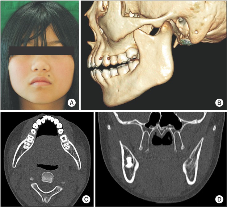
Fig. 5 At 11-months follow-up. A. Clinical photograph shows symmetrical face contour. B, C. Disappearing of newly formed bone and sclerotic change on mandibular ramus and condyle. D. Extraction socket of the third molar completely healing.
Cited by 1 articles
-
Radiographic patterns of periosteal bone reactions associated with endodontic lesions
Poorya Jalali, Jessica Riccobono, Robert A. Augsburger, Mehrnaz Tahmasbi-Arashlow
Restor Dent Endod. 2023;48(3):e23. doi: 10.5395/rde.2023.48.e23.
Reference
-
1. Langlais RP, Langland OE, Nortjé CJ. Diagnostic imaging of the jaws. Baltimore: Williams and Wilkins;1995.2. Neville BW, Damm DD, Allen CM. Oral and maxillofacial pathology. 2nd ed. Philadelphia: Saunders;2002.3. Zand V, Lotfi M, Vosoughhosseini S. Proliferative periostitis: a case report. J Endod. 2008; 34:481–483. PMID: 18358903.
Article4. Jacobson HL, Baumgartner JC, Marshall JG, Beeler WJ. Proliferative periostitis of Garré: report of a case. Oral Surg Oral Med Oral Pathol Oral Radiol Endod. 2002; 94:111–114. PMID: 12193904.
Article5. Ebihara A, Yoshioka T, Suda H. Garrè's osteomyelitis managed by root canal treatment of a mandibular second molar: incorporation of computed tomography with 3D reconstruction in the diagnosis and monitoring of the disease. Int Endod J. 2005; 38:255–261. PMID: 15810976.6. Tong AC, Ng IO, Yeung KM. Osteomyelitis with proliferative periostitis: an unusual case. Oral Surg Oral Med Oral Pathol Oral Radiol Endod. 2006; 102:e14–e19. PMID: 17052617.
Article7. Nortjé CJ, Wood RE, Grotepass F. Periostitis ossificans versus Garrè's osteomyelitis Part II: radiologic analysis of 93 cases in the jaws. Oral Surg Oral Med Oral Pathol. 1988; 66:249–260. PMID: 3140161.8. Wood NK, Goaz PW. Differential diagnosis of oral and maxillofacial lesions. 5nd ed. St Louis: Mosby;1997.
- Full Text Links
-
- Actions
-
Cited
- CITED
-
- Close
- Share
-
- Similar articles
-
- Conservative Treatment of Chronic Suppurative Osteomyelitis on Mandibular Body to Condyle Area: A Case Report
- Postoperative positional change of condyle after bilateral sagittal split ramus osteotomy associated with mandibular asymmetry
- Correction of Facial Asymmetry Using Costochondral Graft and Orthognathic Surgery in Hemifacial Microsomia Patient: Case Report
- Overview of Mandibular Condyle Fracture
- The Change of the Temporomandibular Joint after Experimental Distraction of Mandibular Ramus in Rabbit
