Korean J Ophthalmol.  2015 Dec;29(6):433-434. 10.3341/kjo.2015.29.6.433.

Lacrimal Intrasaccal Cyst

Affiliations
  • 1Department of Ophthalmology, Hanyang University Hospital, Hanyang University College of Medicine, Seoul, Korea.
  • 2Mettapracharak Eye Center, Mettapracharak Hospital, Nakhon Pathom, Thailand.
  • 3Department of Ophthalmology, Samsung Medical Center, Sungkyunkwan University School of Medicine, Seoul, Korea. eyeminded@skku.edu

Abstract

No abstract available.


MeSH Terms

Adult
Cysts/*diagnostic imaging/surgery
Dacryocystorhinostomy
Female
Humans
Lacrimal Apparatus/*diagnostic imaging/surgery
Lacrimal Duct Obstruction/*diagnostic imaging/therapy
Male
Tomography, X-Ray Computed

Figure

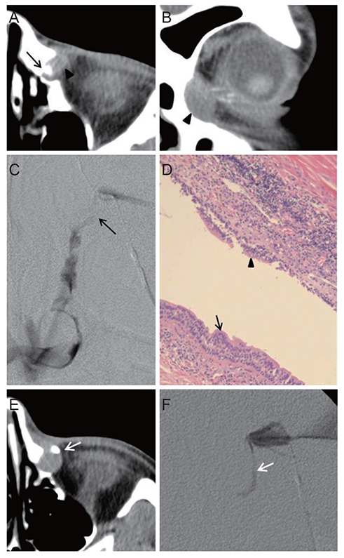
  • Fig. 1 (A,B) Computed tomographic dacryocystography (DCG) demonstrates an ovoid soft tissue mass (arrowhead) anterior to the lacrimal passage (arrow) in the lacrimal sac fossa (case 1). (C) DCG reveals a filling defect in the lacrimal sac without passage disturbance of the dye (arrow) (case 1). (D) The wall of the cystic mass is lined with pseudostratified columnar and cuboidal epithelium (arrowhead), which is infiltrated with inflammatory cells, sharing with the normal lacrimal sac (arrow) (H&E, ×100; case 1). (E) A soft tissue mass is shown in the lacrimal sac fossa with bony remodeling. The dacryocystographic dye (arrow) is deviated anteriolaterally by the cystic mass in the lacrimal sac (case 2). (F) DCG showed a deviated lacrimal sac (arrow) by a space-occupying filling defect on the left side (case 2).


Reference

1. Hosal BM, Hurwitz JJ, Howarth DJ. Orbital cyst of lacrimal sac derivation. Eur J Ophthalmol. 1996; 6:279–283.
2. Hornblass A, Gabry JB. Diagnosis and treatment of lacrimal sac cysts. Ophthalmology. 1979; 86:1655–1661.
3. Sevel D. Development and congenital abnormalities of the nasolacrimal apparatus. J Pediatr Ophthalmol Strabismus. 1981; 18:13–19.
4. Takahashi Y, Nakano T, Asamoto K, et al. Lacrimal sac septum. Orbit. 2012; 31:416–417.
5. Mansour K, Versteegh M, Janssen A, Blanksma L. Epiphora due to compression of the lacrimal sac by a supernumerary blind sac. Orbit. 2002; 21:43–47.
Full Text Links
  • KJO
Actions
Cited
CITED
export Copy
Close
Share
  • Twitter
  • Facebook
Similar articles
Copyright © 2024 by Korean Association of Medical Journal Editors. All rights reserved.     E-mail: koreamed@kamje.or.kr