Korean J Ophthalmol.
2015 Oct;29(5):331-335. 10.3341/kjo.2015.29.5.331.
Structural and Functional Outcomes in Chronic Central Serous Chorioretinopathy Treated with Photodynamic Therapy
- Affiliations
-
- 1Ophthalmology Service, Hospital Universitario La Paz, Madrid, Spain. pinocidad@hotmail.com
- KMID: 2363807
- DOI: http://doi.org/10.3341/kjo.2015.29.5.331
Abstract
- PURPOSE
To study the retinal pigment epithelium (RPE) and retinal alterations in chronic central serous chorioretinopathy treated with photodynamic therapy, and its correlation with functional parameters such as best-corrected visual acuity (BCVA) and contrast sensitivity (CS).
METHODS
Retrospective, noncomparative, consecutive evaluation by optical coherence tomography and its correlation with BCVA and CS in 31 eyes of 26 patients.
RESULTS
In all affected patients, 88.5% were male with a mean age of 42.9 years. The right eye was involved in 64.5% of cases, bilateral in 19% and 73.9% were hyperopic (spherical refraction between 0 and +5.0 diopters). Of these cases, 51.5% had peri-RPE abnormalities, 17.3% hyperreflective substances at RPE, 19.4% RPE atrophy, 55.3% foveolar atrophy, 3.1% pigment epithelial detachment, 5.2% subretinal fluid persistence, 8.3% fibrin deposits, 68.4% photoreceptor inner and outer segment line interruption and 31.1% external limiting membrane interruption.
CONCLUSIONS
Time evolution and number of outbreaks were related to the decrease in foveal and chorodial thickness and in those with worse BCVA and CS. RPE abnormalities and atrophy were related to the age of onset of symptoms. Photoreceptor elongation has been correlated with poor BCVA and inner and outer segment line destructuring and interruption with poor CS.
Keyword
MeSH Terms
-
Adult
Central Serous Chorioretinopathy/diagnosis/*drug therapy/physiopathology
Chronic Disease
Female
Fluorescein Angiography
Follow-Up Studies
Fundus Oculi
Humans
Male
Middle Aged
Photochemotherapy/*methods
Photosensitizing Agents/administration & dosage
Porphyrins/*administration & dosage
Retina/*diagnostic imaging/drug effects/physiopathology
Retrospective Studies
Tomography, Optical Coherence
Treatment Outcome
*Visual Acuity
Photosensitizing Agents
Porphyrins
Figure
-
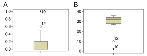
Fig. 1 (A) Affected eye best-corrected visual acuity (logarithm of the minimum angle of resolution). (B) Affected eye contrast sensitivity.
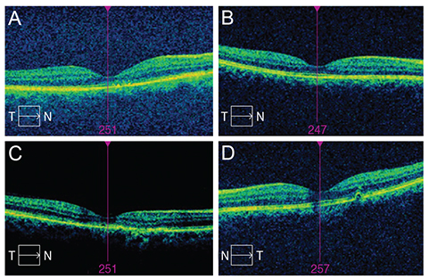
Fig. 2 (A) Case A with hyper-reflective substances visualized by optical coherence tomography (OCT). (B) Case B shows anatomic success objectified by OCT. (C) Case C, OCT demonstrates foveolar atrophy. (D) Case D, pigment epithelial detachment and inner and outer segment line interruption. T = temporal; N = nasal.
Reference
-
1. Fujita K, Shinoda K, Matsumoto CS, et al. Microperimetric evaluation of chronic central serous chorioretinopathy after half-dose photodynamic therapy. Clin Ophthalmol. 2012; 6:1681–1687.2. Song IS, Shin YU, Lee BR. Time-periodic characteristics in the morphology of idiopathic central serous chorioretinopathy evaluated by volume scan using spectral-domain optical coherence tomography. Am J Ophthalmol. 2012; 154:366–375.e4.3. Maruko I, Iida T, Sugano Y, et al. Subfoveal choroidal thickness after treatment of central serous chorioretinopathy. Ophthalmology. 2010; 117:1792–1799.4. Maruko I, Iida T, Sugano Y, et al. Subfoveal choroidal thickness in fellow eyes of patients with central serous chorioretinopathy. Retina. 2011; 31:1603–1608.5. Matsumoto H, Kishi S, Sato T, Mukai R. Fundus autofluorescence of elongated photoreceptor outer segments in central serous chorioretinopathy. Am J Ophthalmol. 2011; 151:617–623.e1.6. Maruko I, Iida T, Sugano Y, et al. One-year choroidal thickness results after photodynamic therapy for central serous chorioretinopathy. Retina. 2011; 31:1921–1927.7. Fraunfelder FW, Fraunfelder FT. Cent ral serous chorioretinopathy associated with sildenafil. Retina. 2008; 28:606–609.8. Asensio-Sanchez VM, Rodriguez-Delgado B, Garcia-Herrero E, et al. Central serous chorioretinopathy as an extradigestive manifestation of Helicobacter pylori gastric infection. Arch Soc Esp Oftalmol. 2008; 83:177–182.9. Kim YY, Flaxel CJ. Factors influencing the visual acuity of chronic central serous chorioretinopathy. Korean J Ophthalmol. 2011; 25:90–97.10. Shinojima A, Kawamura A, Mori R, et al. Detection of morphologic alterations by spectral-domain optical coherence tomography before and af ter half-dose verteporfin photodynamic therapy in chronic central serous chorioretinopathy. Retina. 2011; 31:1912–1920.11. Jirarattanasopa P, Ooto S, Tsujikawa A, et al. Assessment of macular choroidal thickness by optical coherence tomography and angiographic changes in central serous chorioretinopathy. Ophthalmology. 2012; 119:1666–1678.12. Chan WM, Lam DS, Lai TY, et al. Choroidal vascular remodelling in central serous chorioretinopathy after indocyanine green guided photodynamic therapy with verteporfin: a novel treatment at the primary disease level. Br J Ophthalmol. 2003; 87:1453–1458.13. Fujita K, Shinoda K, Imamura Y, et al. Correlation of integrity of cone outer segment tips line with retinal sensitivity after half-dose photodynamic therapy for chronic central serous chorioretinopathy. Am J Ophthalmol. 2012; 154:579–585.14. Vasconcelos H, Marques I, Santos AR, et al. Long-term chorioretinal changes after photodynamic therapy for chronic central serous chorioretinopathy. Graefes Arch Clin Exp Ophthalmol. 2013; 251:1697–1705.15. Gonzalez LS, Abreu Gonzalez R, Alonso Plasencia M, Abreu Reyes P. Normal volume and thickness in spectral-domain OCT: literature revision. Arch Soc Canar Oftalmol. 2012; 23:5.16. Photodynamic therapy of subfoveal choroidal neovascularization in age-related macular degeneration with verteporfin: one-year results of 2 randomized clinical trials. TAP report. Treatment of age-related macular degeneration with photodynamic therapy (TAP) Study Group. Arch Ophthalmol. 1999; 117:1329–1345.17. Kim HC, Cho WB, Chung H. Morphologic changes in acute central serous chorioretinopathy using spectral domain optical coherence tomography. Korean J Ophthalmol. 2012; 26:347–354.18. Tsakonas GD, Kotsolis AI, Koutsandrea C, et al. Multiple spots of photodynamic therapy for the treatment of severe chronic central serous chorioretinopathy. Clin Ophthalmol. 2012; 6:1639–1644.19. Castro-Correia J, Coutinho MF, Rosas V, Maia J. Long-term follow-up of central serous retinopathy in 150 patients. Doc Ophthalmol. 1992; 81:379–386.20. Iida T, Yannuzzi LA, Spaide RF, et al. Cystoid macular degeneration in chronic central serous chorioretinopathy. Retina. 2003; 23:1–7.21. Weingessel B, Mihaltz K, Vecsei-Marlovits PV. Predictors of 1-year visual outcome in OCT analysis comparing ranibizumab monotherapy versus combination therapy with PDT in exsudative age-related macular degeneration. Wien Klin Wochenschr. 2015; 03. 19. DOI: 10.1007/s00508-015-0772-0.22. Ratanasukon M, Thongthong K, Bhurayanontachai P, Jirarattanasopa P. Photoreceptor disruption in central serous chorioretinopathy treated by half-dose photodynamic therapy. Clin Ophthalmol. 2013; 7:87–92.23. Wirostko WJ, Nordgren RN, Dubis AM. Topographical choroidal thickness change following pdt for csc: an oct case report. J Ophthalmol. 2012; 2012:347206.
- Full Text Links
-
- Actions
-
Cited
- CITED
-
- Close
- Share
-
- Similar articles
-
- Photodynamic Therapy With Verteporfin using Half Fluence for Chronic Central Serous Chorioretinopathy
- Review and update for central serous chorioretinopathy
- Photodynamic Therapy with Vertepofin for Short Time for Chronic Central Serous Chorioretinopathy
- Chronic Central Serous Chorioretinopathy: Photodynamic Therapy
- Laser-Induced Choroidal Neovascularization in Central Serous Chorioretinopathy: 4 Cases
