J Korean Ophthalmol Soc.
2012 Jun;53(6):886-894.
Laser-Induced Choroidal Neovascularization in Central Serous Chorioretinopathy: 4 Cases
- Affiliations
-
- 1Department of Ophthalmology, Konkuk University Medical Center, Konkuk University School of Medicine, Seoul, Korea. eyekim@kuh.ac.kr
Abstract
- PURPOSE
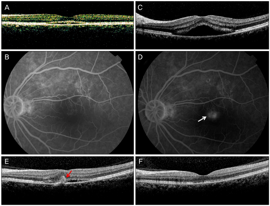
To report the choroidal neovascularization of 4 patients in central serous chorioretinopathy treated with laser treatment.
CASE SUMMARY
We reported 4 patients with central serous chorioretinopathy. Three patients were treated with focal laser photocoagulation, and 1 patient with photodynamic treatment. All patients newly developed choroidal neovascularization (CNV) after treatment at the area of laser treatment. Three patients received intravitreal anti-VEGF injection and 1 patient received argon laser photocoagulation for the treatment of CNV. After the treatment, the CNV was resolved with improved visual acuity.
CONCLUSIONS
Laser treatment may be efficacious tool to achieve rapid resolution in central serous chorioretinopathy until now. However, more caution at the time of treatment and close follow-up after treatment are warranted.
Keyword
MeSH Terms
Figure
Reference
-
1. Gass JD, Norton EW, Justice J Jr. Serous detachment of the retinal pigment epithelium. Trans Am Acad Ophthalmol Otolaryngol. 1966. 70:990–1015.2. Yannuzzi LA. Type-A behavior and central serous chorioretinopathy. Retina. 1987. 7:111–131.3. Spaide RF, Campeas L, Haas A, et al. Central serous chorioretinopathy in younger and older adults. Ophthalmology. 1996. 103:2070–2079.4. Huang WC, Chen WL, Tsai YY, et al. Intravitreal bevacizumab for treatment of chronic central serous chorioretinopathy. Eye (Lond). 2009. 23:488–489.5. Niegel MF, Schrage NF, Christmann S, Degenring RF. [Intravitreal bevacizumab for chronic central serous chorioretinopathy]. Ophthalmologe. 2008. 105:943–945.6. Leaver P, Williams C. Argon Laser photocoagulation in the treatment of central serous retinopathy. Br J Ophthalmol. 1979. 63:674–677.7. Novak MA, Singerman LJ, Rice TA. Krypton and argon laser photocoagulation for central serous chorioretinopathy. Retina. 1987. 7:162–169.8. Chan WM, Lam DS, Lai TY, et al. Choroidal vascular remodelling in central serous chorioretinopathy after indocyanine green guided photodynamic therapy with verteporfin: a novel treatment at the primary disease level. Br J Ophthalmol. 2003. 87:1453–1458.9. Schatz H, Yannuzzi LA, Gitter KA. Subretinal neovascularization following argon laser photocoagulation treatment for central serouschorioretinopathy: complication or misdiagnosis? Trans Sect Ophthalmol Am Acad OPhthalmol Otolaryngol. 1977. 83:893–906.10. Watzke RC, Burton TC, Woolson RF. Direct and indirect laser photocoagulation of central serous choroidopathy. Am J Ophthalmol. 1979. 88:914–918.11. Ha TW, Ham DI, Kang SW. Management of choroidal neovascularization following laser photocoagulation for central serous chorioretinopathy. Korean J Ophthalmol. 2002. 16:88–92.12. Watzke RC, Burton TC, Leaverton PE. Ruby laser photocoagulation therapy of central serous retinopathy. I. A controlled clinical study. II. Factors affecting prognosis. Trans Am Acad Ophthalmol Otolaryngol. 1974. 78:OP205–OP211.13. Klein ML, Van Buskirk EM, Friedman E, et al. Experience with nontreatment of central serous choroidopathy. Arch Ophthalmol. 1974. 91:247–250.14. Bandello F, Virgili G, Lanzetta P, et al. [ICG angiography and retinal pigment epithelial decompensation (CRSC and epitheliopathy)]. J Fr Ophtalmol. 2001. 24:448–451.15. Maumenee AE. Macular diseases: clinical manifestations. Trans Am Acad Ophthalmol Otolaryngol. 1965. 69:605–613.16. Spitznas M. Pathogenesis of central serous retinopathy: a new working hypothesis. Graefes Arch Clin Exp Ophthalmol. 1986. 224:321–324.17. Miller S, Farber D. Cyclic AMP modulation of ion transport across frog retinal pigment epithelium. Measurements in the short-circuit state. J Gen Physiol. 1984. 83:853–874.18. Schlingemann RO. Role of growth factors and the wound healing response in age-related macular degeneration. Graefes Arch Clin Exp Ophthalmol. 2004. 242:91–101.19. Kliffen M, Sharma HS, Mooy CM, et al. Increased expression of angiogenic growth factors in age-related maculopathy. Br J Ophthalmol. 1997. 81:154–162.20. Frank RN, Amin RH, Eliott D, et al. Basic fibroblast growth factor and vascular endothelial growth factor are present in epiretinal and choroidal neovascular membranes. Am J Ophthalmol. 1996. 122:393–403.21. Taban M, Boyer DS, Thomas EL, Taban M. Chronic central serous chorioretinopathy: Photodynamic therapy. Am J Ophthalmol. 2004. 137:1073–1080.22. Canakis C, Livir-Rallatos C, Panayiotis Z, et al. Ocular photodynamic therapy for serous macular detachment in the diffuse retinal pigment epitheliopathy variant of idiopathic central serous chorioretinopathy. Am J Ophthalmol. 2003. 136:750–752.23. Schmidt-Erfurth U, Laqua H, Schlötzer-Schrehard U, et al. Histopathological changes following photodynamic therapy in human eyes. Arch Ophthalmol. 2002. 120:835–844.24. Cardillo Piccolino F, Eandi CM, Ventre L, et al. Photodynamic therapy for chronic central serous chorioretinopathy. Retina. 2003. 23:752–763.25. Olivier S, Harissi-Dagher M, Sébag M. Photodynamic therapy for chronic serous detachment of the retinal pigment epithelium in a young patient. Can J Ophthalmol. 2005. 40:214–216.
- Full Text Links
- Actions
-
Cited
- CITED
-
- Close
- Share
- Similar articles
-
- Management of choroidal neovascularization following laser photocoagulation for central serous chorioretinopathy
- Intravitreal Bevacizumab Injection for Choroidal Neovascularization Secondary to Laser Photocoagulation for Central Serous Chorioretinopathy
- The Optical Coherence Tomography Analysis of Hyperfluorescence by Scanning Laser Ophthalmoscope in Central Serous Chorioretinopathy
- Choroidal Ne ovascularization in Patients with Chronic Central Serous Chorioretinopathy
- A Case of Focal Choroidal Excavation Associated with Chronic Central Serous Chorioretinopathy


