Addition of Digital Breast Tomosynthesis to Full-Field Digital Mammography in the Diagnostic Setting: Additional Value and Cancer Detectability
- Affiliations
-
- 1Department of Radiology, Kyung Hee University Hospital, Kyung Hee University College of Medicine, Seoul, Korea.
- 2Department of Radiology, Seoul National University Hospital, Seoul National University College of Medicine, Seoul, Korea. imchangjm@gmail.com
- 3Department of Radiology, Human Medical Imaging & Intervention Center, Seoul, Korea.
- 4Department of Radiology, Kyungpook National University Medical Center, Daegu, Korea.
- 5Department of Radiology, Good Morning Hospital, Pyeongtaek, Korea.
- 6Department of Radiology, Hanyang University College of Medicine, Seoul, Korea.
- KMID: 2362931
- DOI: http://doi.org/10.4048/jbc.2016.19.4.438
Abstract
- PURPOSE
The purpose of this study was to assess the value of adding digital breast tomosynthesis (DBT) to full-field digital mammography (FFDM) in the diagnostic workup of breast cancer and to determine which lesion variables affect cancer detectability in the combined modality.
METHODS
Between March and May 2012, paired FFDM and DBT images were obtained from 203 women as part of a diagnostic workup for breast cancer. Images from FFDM alone, DBT alone, and DBT combined with FFDM were reviewed in separate sessions by six blinded readers. Jackknife alternative free-response receiver operating characteristic (JAFROC) figure of merit (FOM), sensitivity, and specificity were compared between the modalities. Lesion characteristics affecting the cancer detection rate when using the combined modality were also analyzed.
RESULTS
Among the 203 women, 126 women had a total of 129 malignancies and 77 women had total of 77 benign lesions. The overall JAFROC FOM of the combined modality was higher than that of FFDM alone (0.827 vs. 0.775, p<0.001) and that of DBT alone was higher than that of FFDM alone (0.807 vs. 0.775, p=0.027). The overall sensitivity of the combined modality was higher than that of FFDM alone (80.0% vs. 73.2%, p<0.001) and that of DBT alone was higher than that of FFDM alone (78.3% vs. 73.2%, p=0.007). Compared to FFDM alone, the combined modality detected an additional 48 cancers. Using the combined modality, the presence of masses or microcalcifications was significantly associated with the cancer detection rate (p<0.001).
CONCLUSION
The combination of DBT with FFDM results in a higher diagnostic yield than FFDM alone. Additionally, DBT alone performs better than FFDM alone. However, even when DBT is combined with FFDM, breast cancers with no discernible masses and those lacking calcifications are difficult to detect.
MeSH Terms
Figure
-
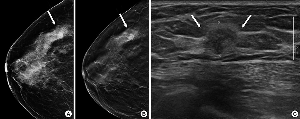
Figure 1 Invasive ductal carcinoma in a 50-year-old woman. (A) Craniocaudal view of full-field digital mammography (FFDM) demonstrates a mass largely obscured by overlying breast tissue (arrow) which was misinterpreted as being negative by four of the six blinded readers. (B) Craniocaudal view of digital breast tomosynthesis (DBT), however, clearly demonstrates the mass (arrow) and all six readers detected the mass on combined FFDM and DBT. (C) Ultrasonography image shows a 1.5-cm irregular, hypoechoic mass with indistinct margins (arrows).

Figure 2 Invasive ductal carcinoma in a 47-year-old woman. (A) Craniocaudal view of full-field digital mammography shows a heterogeneously dense breast tissue which was interpreted as negative by six blinded readers. (B) Craniocaudal view of digital breast tomosynthesis also shows heterogeneously dense breast tissue at the same location. (C) Ultrasonography image shows a 1.1-cm irregular, hypoechoic mass with indistinct margins (arrows).
Reference
-
1. Youlden DR, Cramb SM, Dunn NA, Muller JM, Pyke CM, Baade PD. The descriptive epidemiology of female breast cancer: an international comparison of screening, incidence, survival and mortality. Cancer Epidemiol. 2012; 36:237–248.
Article2. Paap E, Holland R, den Heeten GJ, van Schoor G, Botterweck AA, Verbeek AL, et al. A remarkable reduction of breast cancer deaths in screened versus unscreened women: a case-referent study. Cancer Causes Control. 2010; 21:1569–1573.
Article3. Tabár L, Vitak B, Chen TH, Yen AM, Cohen A, Tot T, et al. Swedish two-county trial: impact of mammographic screening on breast cancer mortality during 3 decades. Radiology. 2011; 260:658–663.
Article4. Pisano ED, Gatsonis C, Hendrick E, Yaffe M, Baum JK, Acharyya S, et al. Diagnostic performance of digital versus film mammography for breast-cancer screening. N Engl J Med. 2005; 353:1773–1783.
Article5. Boyd NF, Guo H, Martin LJ, Sun L, Stone J, Fishell E, et al. Mammographic density and the risk and detection of breast cancer. N Engl J Med. 2007; 356:227–236.
Article6. Carney PA, Miglioretti DL, Yankaskas BC, Kerlikowske K, Rosenberg R, Rutter CM, et al. Individual and combined effects of age, breast density, and hormone replacement therapy use on the accuracy of screening mammography. Ann Intern Med. 2003; 138:168–175.
Article7. Gur D, Abrams GS, Chough DM, Ganott MA, Hakim CM, Perrin RL, et al. Digital breast tomosynthesis: observer performance study. AJR Am J Roentgenol. 2009; 193:586–591.
Article8. Poplack SP, Tosteson TD, Kogel CA, Nagy HM. Digital breast tomosynthesis: initial experience in 98 women with abnormal digital screening mammography. AJR Am J Roentgenol. 2007; 189:616–623.
Article9. Good WF, Abrams GS, Catullo VJ, Chough DM, Ganott MA, Hakim CM, et al. Digital breast tomosynthesis: a pilot observer study. AJR Am J Roentgenol. 2008; 190:865–869.
Article10. Andersson I, Ikeda DM, Zackrisson S, Ruschin M, Svahn T, Timberg P, et al. Breast tomosynthesis and digital mammography: a comparison of breast cancer visibility and BIRADS classification in a population of cancers with subtle mammographic findings. Eur Radiol. 2008; 18:2817–2825.
Article11. Teertstra HJ, Loo CE, van den Bosch MA, van Tinteren H, Rutgers EJ, Muller SH, et al. Breast tomosynthesis in clinical practice: initial results. Eur Radiol. 2010; 20:16–24.
Article12. Rafferty EA, Park JM, Philpotts LE, Poplack SP, Sumkin JH, Halpern EF, et al. Assessing radiologist performance using combined digital mammography and breast tomosynthesis compared with digital mammography alone: results of a multicenter, multireader trial. Radiology. 2013; 266:104–113.
Article13. Skaane P, Gullien R, Bjørndal H, Eben EB, Ekseth U, Haakenaasen U, et al. Digital breast tomosynthesis (DBT): initial experience in a clinical setting. Acta Radiol. 2012; 53:524–529.
Article14. Bernardi D, Ciatto S, Pellegrini M, Tuttobene P, Fanto' C, Valentini M, et al. Prospective study of breast tomosynthesis as a triage to assessment in screening. Breast Cancer Res Treat. 2012; 133:267–271.
Article15. Skaane P, Bandos AI, Gullien R, Eben EB, Ekseth U, Haakenaasen U, et al. Comparison of digital mammography alone and digital mammography plus tomosynthesis in a population-based screening program. Radiology. 2013; 267:47–56.
Article16. Haas BM, Kalra V, Geisel J, Raghu M, Durand M, Philpotts LE. Comparison of tomosynthesis plus digital mammography and digital mammography alone for breast cancer screening. Radiology. 2013; 269:694–700.
Article17. Friedewald SM, Rafferty EA, Rose SL, Durand MA, Plecha DM, Greenberg JS, et al. Breast cancer screening using tomosynthesis in combination with digital mammography. JAMA. 2014; 311:2499–2507.
Article18. McCarthy AM, Kontos D, Synnestvedt M, Tan KS, Heitjan DF, Schnall M, et al. Screening outcomes following implementation of digital breast tomosynthesis in a general-population screening program. J Natl Cancer Inst. 2014; 106:dju316.
Article19. Gur D, Bandos AI, Rockette HE, Zuley ML, Sumkin JH, Chough DM, et al. Localized detection and classification of abnormalities on FFDM and tomosynthesis examinations rated under an FROC paradigm. AJR Am J Roentgenol. 2011; 196:737–741.
Article20. Ciatto S, Houssami N, Bernardi D, Caumo F, Pellegrini M, Brunelli S, et al. Integration of 3D digital mammography with tomosynthesis for population breast-cancer screening (STORM): a prospective comparison study. Lancet Oncol. 2013; 14:583–589.
Article21. Houssami N, Skaane P. Overview of the evidence on digital breast tomosynthesis in breast cancer detection. Breast. 2013; 22:101–108.
Article22. Chesebro AL, Winkler NS, Birdwell RL, Giess CS. Developing asymmetry at mammography: correlation with US and MR imaging and histopathologic findings. Radiology. 2016; 279:385–394.
Article23. D'Orsi CJ, Sickles EA, Mendelson EB, Morris EA. ACR BI-RADS® Atlas: Breast Imaging and Reporting and Data System. 5th ed. Reston: American College of Radiology;2013.24. Leung JW, Sickles EA. Developing asymmetry identified on mammography: correlation with imaging outcome and pathologic findings. AJR Am J Roentgenol. 2007; 188:667–675.
Article25. Spangler ML, Zuley ML, Sumkin JH, Abrams G, Ganott MA, Hakim C, et al. Detection and classification of calcifications on digital breast tomosynthesis and 2D digital mammography: a comparison. AJR Am J Roentgenol. 2011; 196:320–324.
Article26. Noroozian M, Hadjiiski L, Rahnama-Moghadam S, Klein KA, Jeffries DO, Pinsky RW, et al. Digital breast tomosynthesis is comparable to mammographic spot views for mass characterization. Radiology. 2012; 262:61–68.
Article27. Kopans DB. Digital breast tomosynthesis: a better mammogram. Radiology. 2013; 267:968–969.
Article28. Hooley RJ, Geisel JL, Raghu M, Durand MA, Gross CP, Busch SH, et al. Performance of whole breast ultrasound in women with dense breasts following 3D tomosynthesis mammography. In : The 99th Radiological Society of North America Scientific Assembly and Annual Meeting; 2013. Abstract #SSQ01-06.29. Mariscotti G, Houssami N, Durando M, Bergamasco L, Campanino PP, Ruggieri C, et al. Accuracy of mammography, digital breast tomosynthesis, ultrasound and MR imaging in preoperative assessment of breast cancer. Anticancer Res. 2014; 34:1219–1225.30. Kim SA, Chang JM, Cho N, Yi A, Moon WK. Characterization of breast lesions: comparison of digital breast tomosynthesis and ultrasonography. Korean J Radiol. 2015; 16:229–238.
Article
- Full Text Links
-
- Actions
-
Cited
- CITED
-
- Close
- Share
-
- Similar articles
-
- Digital Mammography
- Clinical Application of Artificial Intelligence in Digital Breast Tomosynthesis
- Digital Breast Tomosynthesis versus MRI as an Adjunct to Full-Field Digital Mammography for Preoperative Evaluation of Breast Cancer according to Mammographic Density
- Mammography-Guided Interventional Procedure
- Factors Affecting Breast Cancer Detectability on Digital Breast Tomosynthesis and Two-Dimensional Digital Mammography in Patients with Dense Breasts
