The Measurement of Normal Talus in Korean Cadaver
- Affiliations
-
- 1Department of Orthopedic Surgery, Inje University Busan Paik Hospital, Busan, Korea. bluewhisle@gmail.com
- 2Department of Orthopedic Surgery, Maryknoll Medical Center, Busan, Korea.
- 3Department of Anesthesiology and Pain Medicine, Inje University Busan Paik Hospital, Busan, Korea.
- KMID: 2362764
- DOI: http://doi.org/10.14193/jkfas.2016.20.4.163
Abstract
- PURPOSE
To investigate the measured values of the talus in Koreans.
MATERIALS AND METHODS
We measured 88 tali from 44 cadavers that have been donated between December 2012 and December 2015. Of the cadavers, 27 were male and 17 were female. Their mean age was 73 years. The length and width of the talus were measured using a digital goniometer and vernier caliper.
RESULTS
The values of cadaveric measurement, mean maximal width and length, width and length of the dome anterior, width and length of the posterior facet, height and length of the trochlear medial facet, and height and length of the trochlear lateral facet were 43.6±2.6 mm, 56.5±3.3 mm, 32.5±2.0 mm, 42.2±2.7 mm, 22.2±2.2 mm, 34.7±2.0 mm, 15.3±1.3 mm, 33.3±2.9 mm, 25.3±3.3 mm, and 30.8±2.4 mm for men and 38.9±1.6 mm, 53.6±2.4 mm, 27.9±2.1 mm, 37.4±3.2 mm, 20.6±0.8 mm, 31.9±1.2 mm, 13.6±2.6 mm, 28.4±2.5mm, 24.9±2.1 mm, and 28.9µ1.4 mm for women, respectively. The size of the talus showed an accuracy of 86% when anteroposterior diameter was greater than 59 mm. A difference in the size of the right and left talus was not observed. The mean inclination and declination angles were 24.4°±4.2° and 28.2°±5.4° for men, and 24.6°±3.6° and 24.7°±6.7° for women (p=0.980, p=0.018), respectively, at least 15°, which showed a big difference for every object up to 37°.
CONCLUSION
This paper, to the best of our knowledge, is the first study to measure the talus in Koreans. There were differences by gender and ethnicity in the in measured talus values. The measurements were smaller than European-Americans and greater than Japanese.
Figure
-

Figure 1. Actual measurement of total width (longitudinal length from lateral process to talus head) and total length (longitudinal length from talus head to lateral tubercle) of talus in cadaver.

Figure 2. Actual measurement of anterior width (longitudinal length from medial edge of talar dome to lateral edge of talar dome surface at the highest) and length (longitudinal length from anterior edge of talar dome to posterior edge of talar dome surface) of talus in cadaver.

Figure 3. Actual measurement of posterior facet width (longitudinal length from medial edge to lateral edge of posterior facet at posterior calcaneal articular facet surface) and length (longitudinal length from anterior edge to posterior edge of posterior facet at posterior calcaneal articular facet surface) of talus in cadaver.
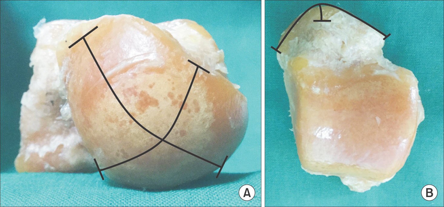
Figure 4. (A, B) Actual measurement of head for navicular width (longitudinal length from medial edge of talar head to lateral edge of talar head) and length (longitudinal length from superior edge of talar head to inferior edge of talar head) of talus in cadaver.
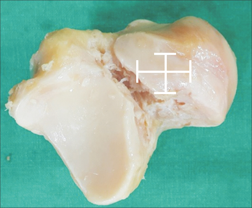
Figure 5. Actual measurement of anterior facet width (longitudinal length from anterior edge of anterior calcaneal articular facet to posterior edge of anterior calcaneal articular facet) and length (longitudinal length from anterior edge of anterior calcaneal articular facet to posterior edge of anterior calcaneal articular facet) of talus in cadaver.
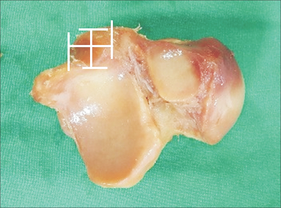
Figure 6. Actual measurement of middle facet width (longitudinal length from medial edge of middle calcaneal articular facet to lateral edge of middle calcaneal articular facet) and length (longitudinal length from anterior edge of middle calcaneal articular facet to posterior edge of middle calcaneal articular facet) of talus in cadaver.

Figure 7. Actual measurement of trochlear medial facet height (longitudinal length from inferior edge of trochlear for medial malleolus to superior edge of trochlear for medial malleolus) and length (longitudinal length from anterior edge of trochlear for medial malleolus to posterior edge of trochlear for medial malleolus) of talus in cadaver.
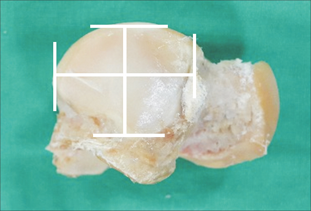
Figure 8. Actual measurement of trochlear lateral facet height (longitudinal length from inferior edge of trochlear for lateral malleolus to superior edge of trochlear for lateral malleolus) and length (longitudinal length from anterior edge of trochlear for lateral malleolus to posterior edge of trochlear for lateral malleolus) of talus in cadaver.
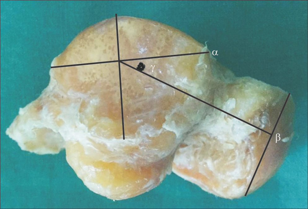
Figure 9. Actual measurement of inclination angle (γ; angle of between longitudinal line [α] of medial trochlear and perpendicular line of talar head [β]) of talus in cadaver.
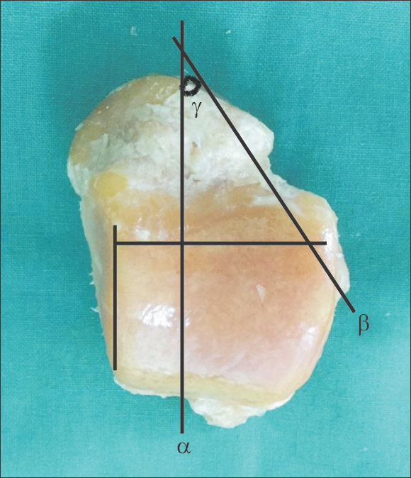
Figure 10. Actual measurement of declination angle (γ; angle of between longitudinal line of superior trochlear [α] and longitudinal line of talar neck [β]) of talus in cadaver.
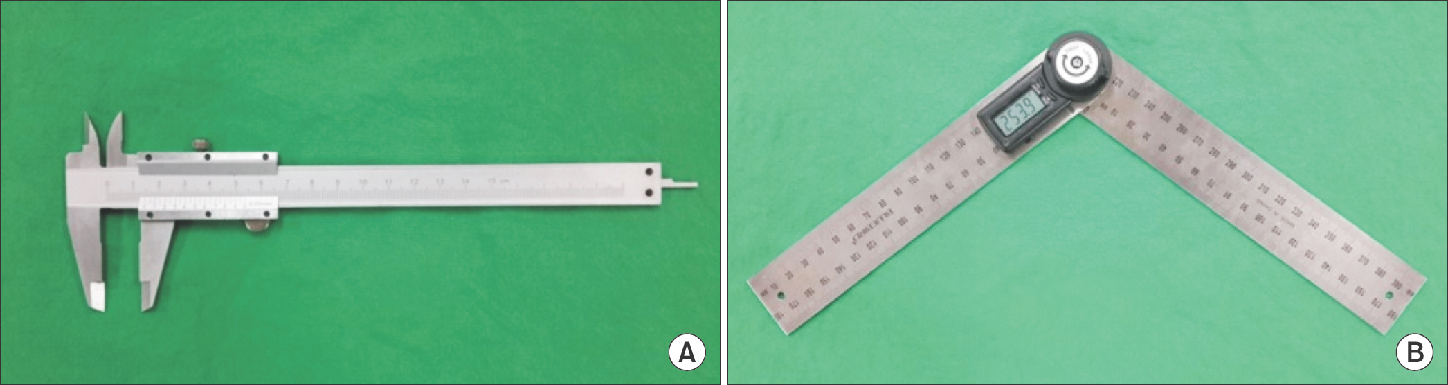
Figure 11. (A) Actual measurements of talus were done in cadaver by vernier calipers (Mitutoyo Philosophy, Kawasaki, Japan). (B) Actual measurements of talus were done in cadaver by digital goniometer (Bluebird, Seoul, Korea).
Reference
-
1.Crossan ET. Fractures of the tarsal scaphoid and of the os calcis. Surg Clin North Am. 1930. 10:1477.2.Art W., Roy WS., Alireza B., John GA., Donald RB. Ankle fractures. Coughlin MJ, Saltzman CL, Anderson RB, editors. editors.Mann’s surgery of the foot and ankle. 9th ed.Philadelphia: Saunders;2014. p.2102.3.Sarrafian SK. Anatomy of the foot and ankle1 Philadelphia: Lip-pincott. 1983.4.Na WC., Lee SH., Lee JY., Lee SJ., Kim B. The result of open reduction and mini-plate fixation for displaced talar neck fracture. J Korean Fract Soc. 2015. 28:215–22.
Article5.Williams PL., Bannister LH., Berry MM., Collins P., Dyson M., Dussek JE, et al. Gray’s anatomy. 38th ed.London: Churchill Livingstone;1999.6.Sakaue K. Sex assessment from the talus and calcaneus of Japa-nese. Bull Natl Mus Nat Sci Ser D. 2011. 37:35–48.7.Gualdi-Russo E. Sex determination from the talus and calcaneus measurements. Forensic Sci Int. 2007. 171:151–6.
Article8.Testut L. Traité d’anatomie humaine. 7th ed.Paris: Doin;1921. p.368.9.Paturet G. Traité d’anatomie humaine. Paris: Masson;1951. p.573.10.Gautham K., Clarista MQ., Sheela N., Vidyashambhava P. Morphometric analysis of the human tali. CIBTech J Surg. 2013. 2:64–8.11.Sewell RBS. A study of the astragalus. J Anat Physiol. 1904. 39:74–88.12.Robinson C., Eisma R., Morgan B., Jeffery A., Graham EA., Black S., Rutty GN. Anthropological measurement of lower limb and foot bones using multi-detector computed tomography. J Forensic Sci. 2008. 53:1289–95.
Article
- Full Text Links
-
- Actions
-
Cited
- CITED
-
- Close
- Share
-
- Similar articles
-
- Treatment for Total Extrusion of the Talus (Missing Talus) using the Sandwich Block Tibiocalcaneonavicular Arthrodesis: A Case Report
- A Case of Atraumatic Aseptic Necrosis of Both Talus: A Case Report
- Micro-Structural Profiles of Trabecular Bone at the Ankle Joint
- Total Dislocation of the Talus: A Case Report of a Three Year Followup
- The Talus-1st Metatarsal Angle, the Talo-Horizontal Angle and Calcaneal Pitch Angle of Young Men in Korea
