Imaging Sci Dent.
2016 Dec;46(4):279-284. 10.5624/isd.2016.46.4.279.
Long-standing chin-augmenting costochondral graft creating a diagnostic challenge: A case report and literature review
- Affiliations
-
- 1Department of Oral and Maxillofacial Diagnostic Sciences, Oral and Maxillofacial Radiology, College of Dentistry, University of Florida, Gainesville, FL, USA. fbadr@dental.ufl.edu
- 2Department of Oral and Maxillofacial Diagnostic Sciences, Oral and Maxillofacial Pathology, College of Dentistry, University of Florida, Gainesville, FL, USA.
- 3Barton Blumberg, DMD Oral and Maxillofacial Surgery Center, The Villages, FL, USA.
- KMID: 2362747
- DOI: http://doi.org/10.5624/isd.2016.46.4.279
Abstract
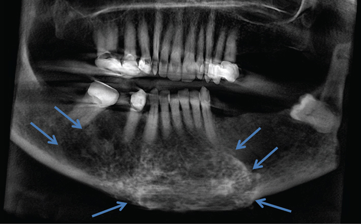
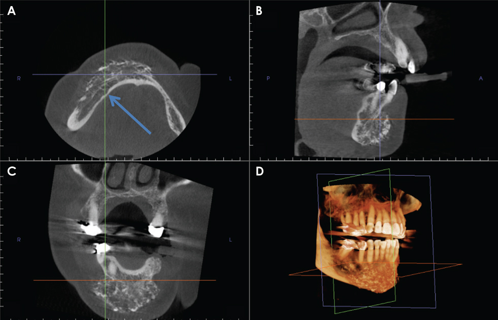
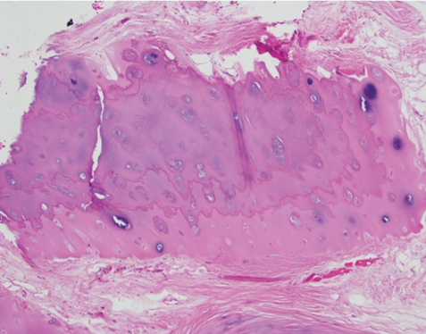
- To our knowledge, the imaging features of costochondral grafts (CCGs) on cone-beam computed tomography (CBCT) have not been documented in the literature. We present the case of a CCG in the facial soft tissue to the anterior mandible, with changes mimicking a cartilaginous neoplasm. This is the first report to describe the CBCT imaging features of a long-standing graft in the anterior mandible. Implants or grafts may be incidental findings on radiographic images made for unrelated purposes. Although most are well-defined and radiographically homogeneous, being of relatively inert non-biological material, immune reactions to some grafts may stimulate alterations in the appearance of surrounding tissues. Biological implants may undergo growth and differentiation, causing their appearance to mimic neoplastic lesions. We present the case of a cosmetic autogenous CCG that posed a diagnostic challenge both radiographically and histopathologically.
MeSH Terms
Figure
Reference
-
1. Rollnick BR, Kaye CI, Nagatoshi K, Hauck W, Martin AO. Oculoauriculovertebral dysplasia and variants: phenotypic characteristics of 294 patients. Am J Med Genet. 1987; 26:361–375.
Article2. Gorlin RJ, Jue KL, Jacobsen U, Goldschmidt E. Oculoauriculovertebral dysplasia. J Pediatr. 1963; 63:991–999.
Article3. Pashayan H, Pinsky L, Fraser FC. Hemifacial microsomia -oculo-auriculo-vertebral dysplasia. A patient with overlapping features. J Med Genet. 1970; 7:185–188.4. Cohen MM Jr, Rollnick BR, Kaye CI. Oculoauriculovertebral spectrum: an updated critique. Cleft Palate J. 1989; 26:276–286.5. Poswillo D. The pathogenesis of the first and second branchial arch syndrome. Oral Surg Oral Med Oral Pathol. 1973; 35:302–328.
Article6. Beleza-Meireles A, Clayton-Smith J, Saraiva JM, Tassabehji M. Oculo-auriculo-vertebral spectrum: a review of the literature and genetic update. J Med Genet. 2014; 51:635–645.
Article7. The free dictionary by FARLEX. Medical-dictionary.thefreedictionary.com [Internet]. cited 2016 April 4. Available from: http://medical-dictionary.thefreedictionary.com.8. Schatz CJ, Ginat DT. Imaging of cosmetic facial implants and grafts. AJNR Am J Neuroradiol. 2013; 34:1674–1681.
Article9. Yang S, Fan H, Du W, Li J, Hu J, Luo E. Overgrowth of costochondral grafts in craniomaxillofacial reconstruction: rare complication and literature review. J Craniomaxillofac Surg. 2015; 43:803–812.
Article10. Eckardt A, Swennen G, Teltzrow T. Melanotic neuroectodermal tumor of infancy involving the mandible: 7-year follow-up after hemimandibulectomy and costochondral graft reconstruction. J Craniofac Surg. 2001; 12:349–354.
Article11. Kaban LB, Perrott DH, Fisher K. A protocol for management of temporomandibular joint ankylosis. J Oral Maxillofac Surg. 1990; 48:1145–1151.
Article12. Kansy K, Mueller AA, Mucke T, Kopp JB, Koersgen F, Wolff KD, et al. Microsurgical reconstruction of the head and neck -current concepts of maxillofacial surgery in Europe. J Craniomaxillofac Surg. 2014; 42:1610–1613.13. Ko EW, Huang CS, Chen YR. Temporomandibular joint reconstruction in children using costochondral grafts. J Oral Maxillofac Surg. 1999; 57:789–798.
Article14. Nelson CL, Buttrum JD. Costochondral grafting for posttraumatic temporomandibular joint reconstruction: a review of six cases. J Oral Maxillofac Surg. 1989; 47:1030–1036.
Article15. Obeid G, Guttenberg SA, Connole PW. Costochondral grafting in condylar replacement and mandibular reconstruction. J Oral Maxillofac Surg. 1988; 46:177–182.
Article16. Fukuta K, Jackson IT, Topf JS. Facial lawn mower injury treated by a vascularized costochondral graft. J Oral Maxillofac Surg. 1992; 50:194–198.
Article17. Raustia A, Pernu H, Pyhtinen J, Oikarinen K. Clinical and computed tomographic findings in costochondral grafts replacing the mandibular condyle. J Oral Maxillofac Surg. 1996; 54:1393–1400.
Article18. Samman N, Cheung LK, Tideman H. Overgrowth of a costochondral graft in an adult male. Int J Oral Maxillofac Surg. 1995; 24:333–335.
Article19. Mallya SM, Lurie AG. Panoramic imaging. In : White SC, Pharoah MJ, editors. Oral radiology: principles and interpretation. 7th ed. St. Louis, Mo.: Mosby/Elsevier;2014. p. 166–184.20. Jang HW, Kim NK, Lee WS, Kim HJ, Cha IH, Nam W. Mandibular condyle and infratemporal fossa reconstruction using vascularized costochondral and calvarial bone grafts. J Korean Assoc Oral Maxillofac Surg. 2014; 40:83–86.
Article21. Karacaoğlan N, Akbas H, Eroğlu L, Ioncesu L. Chin augmentation using diced cartilage. Eur J Plast Surg. 1998; 21:254–256.
Article22. Loftus MJ, Bennett JA, Fantasia JE. Osteochondroma of the mandibular condyles. Report of three cases and review of the literature. Oral Surg Oral Med Oral Pathol. 1986; 61:221–226.23. Garrington GE, Collett WK. Chondrosarcoma. II. Chondrosarcoma of the jaws: analysis of 37 cases. J Oral Pathol. 1988; 17:12–20.
Article
- Full Text Links
- Actions
-
Cited
- CITED
-
- Close
- Share
- Similar articles
-
- Temporomandibular joint reconstruction with costochondral graft: case series study
- A case report of hemifacial microsomia
- TEMPOROMANDIBULAR JOINT RECONSTRUCTION USING COSTOCHONDRAL GRAFT: CASE REPORTS
- Correction of Facial Asymmetry Using Costochondral Graft and Orthognathic Surgery in Hemifacial Microsomia Patient: Case Report
- Comparison of Costochondral Graft and Customized Total Joint Reconstruction for Treatments of Temporomandibular Joint Replacement

