J Korean Soc Radiol.
2016 Dec;75(6):471-479. 10.3348/jksr.2016.75.6.471.
Venous Reflux on Contrast-Enhanced Head and Neck Magnetic Resonance Angiography: Analysis of Causative Factors
- Affiliations
-
- 1Department of Radiology, Dongguk University Ilsan Hospital, Goyang, Korea. ejl1048@hanmail.net
- 2Department of Preventive Medicine, Jeju National University School of Medicine, Jeju, Korea.
- KMID: 2360435
- DOI: http://doi.org/10.3348/jksr.2016.75.6.471
Abstract
- PURPOSE
The purpose of this study was to analyze the causative factors of venous reflux on contrast-enhanced head and neck magnetic resonance angiography.
MATERIALS AND METHODS
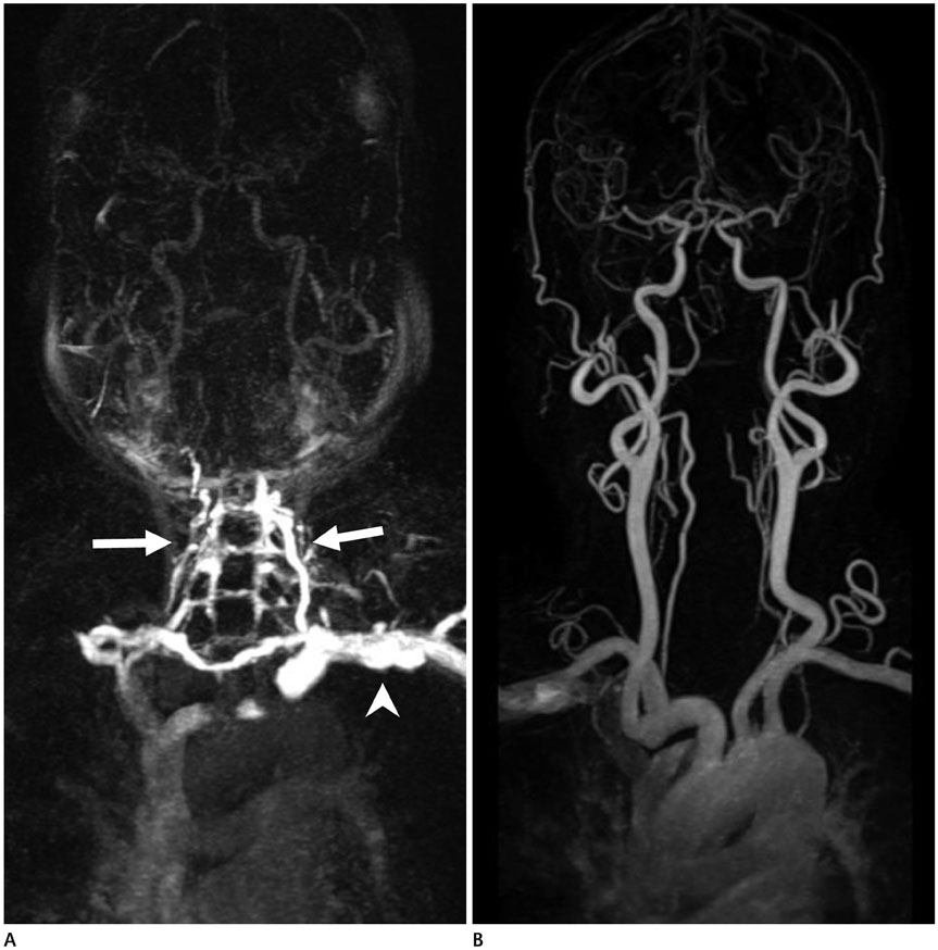
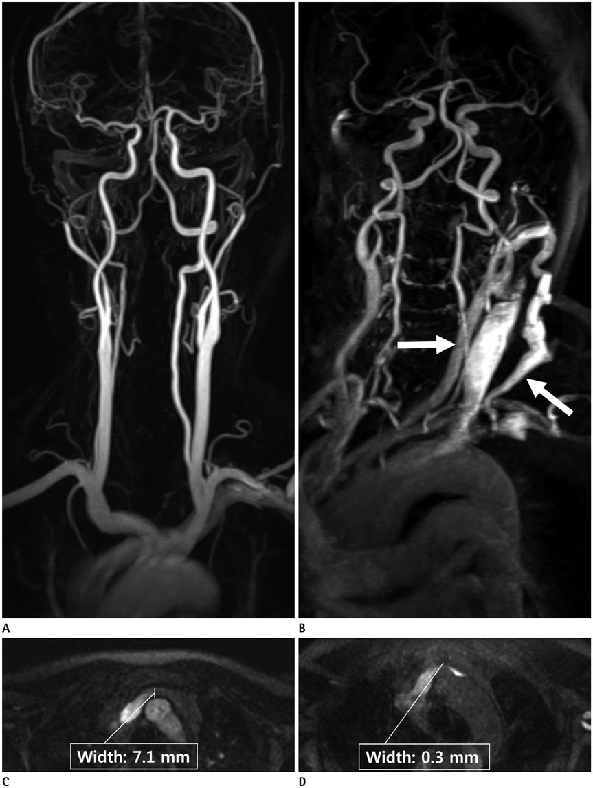
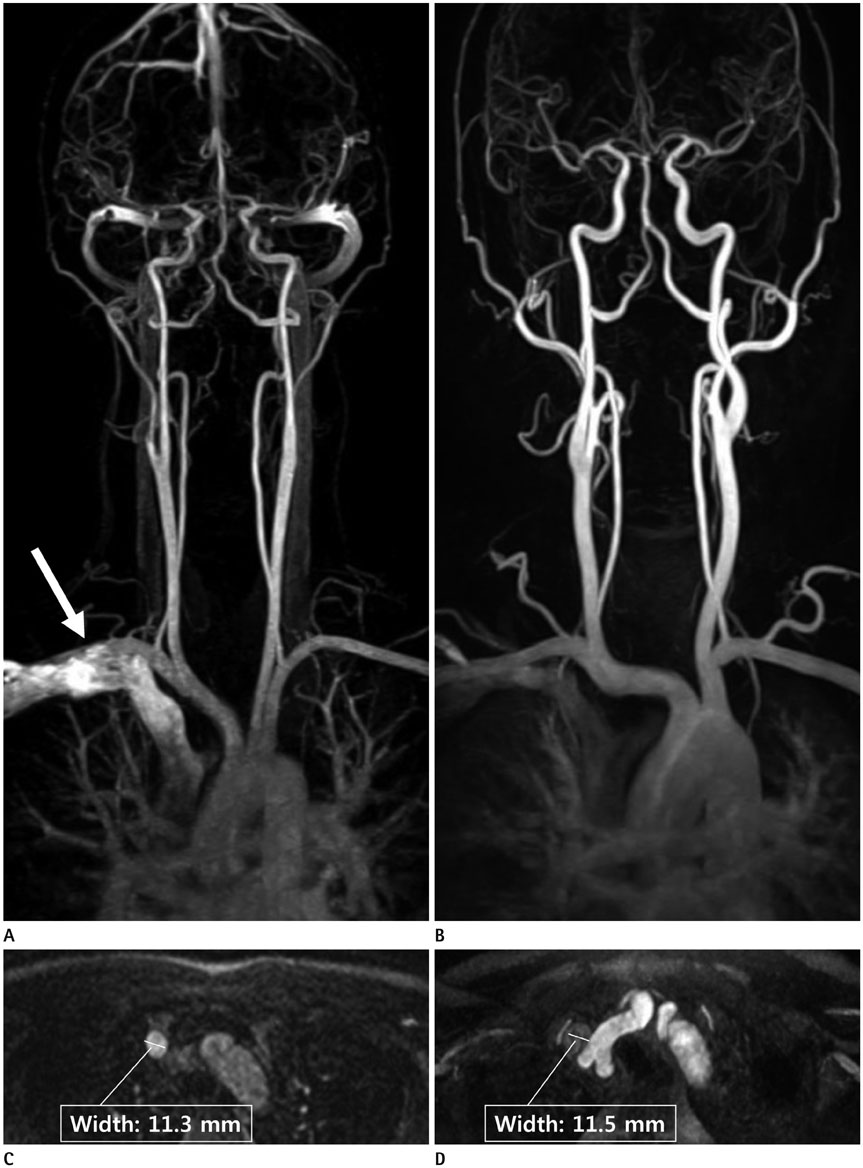
We retrospectively reviewed 150 patients with right-arm injections and 150 patients with left-arm injections. We included the age, gender, body mass index, history of hypertension, and history of diabetes mellitus in the evaluation of all patients. We measured the shortest width of the left or right brachiocephalic vein (BCV), the diameter of the aortic arch, and the distance between the sternum and vertebral body. The relationship between these factors and the venous reflux was analyzed. In patients with venous reflux, we performed qualitative image scoring for suboptimal images.
RESULTS
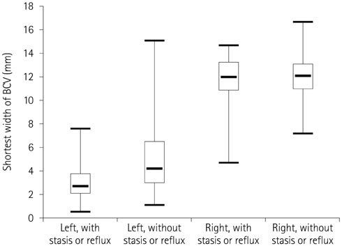
In patients with venous reflux, the image quality of the left-arm injection group was significantly inferior to the image quality of the right-arm injection group. The mean age and the male-to-female ratio of patients with venous reflux were significantly higher than those of patients without venous reflux. In patients receiving the left-arm injection, the mean shortest width of the left BCV was significantly narrower in patients with venous reflux than in patients without venous reflux.
CONCLUSION
A left-arm injection should be avoided, especially in elderly patients, to acquire an optimal image.
MeSH Terms
Figure
Reference
-
1. Prince MR. Gadolinium-enhanced MR aortography. Radiology. 1994; 191:155–164.2. Edelman RR. MR angiography: present and future. AJR Am J Roentgenol. 1993; 161:1–11.3. Cloft HJ, Murphy KJ, Prince MR, Brunberg JA. 3D gadolinium-enhanced MR angiography of the carotid arteries. Magn Reson Imaging. 1996; 14:593–600.4. Tanaka T, Uemura K, Takahashi M, Takehara S, Fukaya T, Tokuyama T, et al. Compression of the left brachiocephalic vein: cause of high signal intensity of the left sigmoid sinus and internal jugular vein on MR images. Radiology. 1993; 188:355–361.5. Hingwala DR, Thomas B, Kesavadas C, Kapilamoorthy TR. Suboptimal contrast opacification of dynamic head and neck MR angiography due to venous stasis and reflux: technical considerations for optimization. AJNR Am J Neuroradiol. 2011; 32:310–314.6. Lee YJ, Chung TS, Joo JY, Chien D, Laub G. Suboptimal contrast-enhanced carotid MR angiography from the left brachiocephalic venous stasis. J Magn Reson Imaging. 1999; 10:503–509.7. You SY, Yoon DY, Choi CS, Chang SK, Yun EJ, Seo YL, et al. Effects of right- versus left-arm injections of contrast material on computed tomography of the head and neck. J Comput Assist Tomogr. 2007; 31:677–681.8. Tseng YC, Hsu HL, Lee TH, Chen CJ. Venous reflux on carotid computed tomography angiography: relationship with left-arm injection. J Comput Assist Tomogr. 2007; 31:360–364.9. Bok B, Marsault C, Aubin ML, Bar D, Aboulker J. Jugular venous reflux in cerebral radionuclide angiography: an explanation. Eur J Nucl Med. 1978; 3:63–65.10. Yeh EL, Pohlman GP, Ruetz PP, Meade RC. Jugular venous reflux in cerebral radionuclide angiography. Radiology. 1976; 118:730–732.11. Demirpolat G, Yüksel M, Kavukçu G, Tuncel D. Carotid CT angiography: comparison of image quality for left versus right arm injections. Diagn Interv Radiol. 2011; 17:195–198.12. Hayt DB, Perez LA. Cervical venous reflux in dynamic brain scintigraphy. J Nucl Med. 1976; 17:9–12.13. Fisher J, Vaghaiwalla F, Tsitlik J, Levin H, Brinker J, Weisfeldt M, et al. Determinants and clinical significance of jugular venous valve competence. Circulation. 1982; 65:188–196.14. Anderhuber F. [Venous valves in the large branches of superior vena cava]. Acta Anat (Basel). 1984; 119:184–192.15. Harmon JV Jr, Edwards WD. Venous valves in subclavian and internal jugular veins. Frequency, position, and structure in 100 autopsy cases. Am J Cardiovasc Pathol. 1987; 1:51–54.16. Fred HL, Wukasch DC, Petrany Z. Transient compression of the left innominate vein. Circulation. 1964; 29:758–761.17. Peart RA, Driedger AA. Effect of obstructed mediastinal venous return on dynamic brain blood flow studies: case report. J Nucl Med. 1975; 16:622–625.18. Silverstein GE, Burke G, Goldberg D, Halko A. Superior vena caval system obstruction caused by benign endothoracic goiter. Dis Chest. 1969; 56:519–523.19. McLintic AJ, Martin W, Tweddel AC. The influence of arm position and cardiac output on bolus clearance from the arm. Anaesthesia. 1992; 47:838–841.20. Hernandez JA, Walser EM, Swischuk LE. Aortosternal venous compression in patients with aberrant right subclavian arteries. AJR Am J Roentgenol. 2005; 184:1434–1436.
- Full Text Links
- Actions
-
Cited
- CITED
-
- Close
- Share
- Similar articles
-
- Resolved Cerebral Venous Hypertension after Angioplasty of Central Venous Stenosis in a Hemodialysis Patient: A Case Report
- Image Findings in Brain Developmental Venous Anomalies
- Evaluation of the Cause of Internal Jugular Vein Obstruction on Head and Neck Contrast Enhanced 3D MR Angiography Using Contrast Enhanced Computed Tomography
- Contrast-Enhanced Magnetic Resonance Angiography: Dose the Test Dose Bolus Represent the Main Dose Bolus Accurately?
- Cerebellar Venous Angioma Confused with Peripheral Vestibulopathy


