Chronic Intussusception Caused by Diffuse Large B-Cell Lymphoma in a 6-Year-Old Girl Presenting with Abdominal Pain and Constipation for 2 Months
- Affiliations
-
- 1Department of Pediatrics, Kyung Hee University Hospital at Gangdong, Seoul, Korea. chsh0414@khu.ac.kr
- 2Department of Surgery, Kyung Hee University Hospital at Gangdong, Seoul, Korea.
- 3Department of Pathology, Kyung Hee University Hospital at Gangdong, Seoul, Korea.
- KMID: 2360060
- DOI: http://doi.org/10.3346/jkms.2016.31.2.321
Abstract
- The classical triad of abdominal pain, vomiting, and bloody stool is absent in chronic intussusception for more than 2 weeks. Here, we report a 6-year-old female with recurrent abdominal pain for 2 months. Ultrasonography of the abdomen revealed an ileocolic-type intussusception. The lesion accompanying the tight fibrous adhesion was treated by resection and ileocolic anastomosis. It was diagnosed as intussusception with diffuse large B-cell lymphoma. A high index of suspicion for abdominal pain in children should result in the correct diagnosis and appropriate management.
Keyword
MeSH Terms
Figure
-
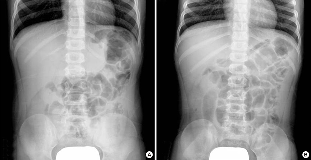
Fig. 1 Plain abdominal radiographs taken in the emergency room 3 days before visiting the outpatient clinic showed mild to moderate ileus without definite mechanical obstruction. (A) Erect view. (B) Supine view.
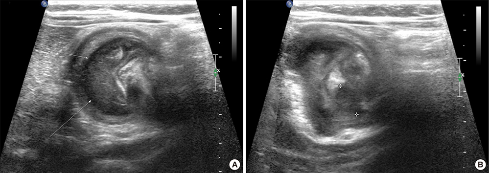
Fig. 2 Abdominal ultrasonography of the right upper abdomen. (A) An ileocolic-type intussusception was found with a crescent-shaped hypoechoic portion (arrows). (B) Enlarged lymph nodes (*) along the intussusceptions.

Fig. 3 Abdominal computed tomography. (A) A 3-cm, crescent-shaped (arrow), low-density lesion at the top of the intussusception. (B) Ileocolic-type intussusception (arrow).
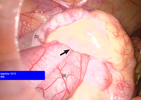
Fig. 4 Laparoscopy view showing the intussusception. Distal ileum (a) is pulled into ascending colon (b), forming ileocolic type intussuception (arrow).

Fig. 5 Gross and microscopic findings of the resected tissue. (A) Gross view of the formalin-fixed hemicolectomy specimen including ileum, cecum and appendix showing an ill-defined ulcerative lesion (arrow), measuring 5.5 × 4.0 cm. (B) The terminal ileum shows ulceration with diffuse tumor cell infiltration (arrow) in the mucosa and proper muscle layer in a low-magnification view (H&E stained, × 20). (C) The tumor cells have large vesicular nuclei with prominent nucleolus and thick nuclear membrane (H&E stained, × 400). (D) The subserosa and serosa of the terminal ileum show fibrosis and chronic inflammatory cell infiltration (arrow) admixed with tumor cell infiltration (Masson trichrome stained, × 20).
Reference
-
1. Malakounides G, Thomas L, Lakhoo K. Just another case of diarrhea and vomiting? Pediatr Emerg Care. 2009; 25:407–410.2. Reijnen JA, Festen C, Joosten HJ. Chronic intussusception in children. Br J Surg. 1989; 76:815–816.3. Singal R, Gupta S, Goel M, Jain P. A rare case of chronic intussusception due to non Hodgkin lymphoma. Acta Gastroenterol Belg. 2012; 75:42–44.4. Yadav K, Patel RV, Mitra SK, Malik AK. Chronic secondary caeco-colic intussusception in a boy associated with primary malignant lymphoma of caecum (a case report). J Postgrad Med. 1986; 32:94–96.5. Shakya VC, Agrawal CS, Koirala R, Khaniya S, Rajbanshi S, Pandey SR, Adhikary S. Intussusception due to non Hodgkin’s lymphoma; different experiences in two children: two case reports. Cases J. 2009; 2:6304.6. Seo HE, Lee JH, Park TI, Lee KS. Small intestinal mucosa associated lymphoid tissue(MALT) lymphoma in a child presenting with recurrent intussusceptions: a case report. Korean J Hematol. 2007; 42:419–422.7. Kim KN, Woo JJ, Bahk YW, Kim SY, Kang YM. High grade MALT lymphoma of the ileum in a child presenting as intussusceptions: a case report. J Korean Radiol Soc. 2002; 46:279–282.
- Full Text Links
-
- Actions
-
Cited
- CITED
-
- Close
- Share
-
- Similar articles
-
- Distal Ileal Lymphoma Presenting Ileocecal Intussusception with Spontaneous Reduction
- A 9-Month-Old Boy with a Recurrent Ileocolic Intussusception Caused by Diffuse Large B Cell Lymphoma: A Case Report
- Chronic Constipation Led to Sigmoid Volvulus in a Child
- A Case of Epstein-Barr Virus-Positive Diffuse Large B-Cell Lymphoma Occurring in Thyroid Gland
- Two cases of cecal lymphoma causing intussusception
