Calcified Pulmonary Nodules Identified in a 350-Year-Old-Joseon Mummy: the First Report on Ancient Pulmonary Tuberculosis from Archaeologically Obtained Pre-modern Korean Samples
- Affiliations
-
- 1Department of Anatomy, Ewha Womans University School of Medicine, Seoul, Korea.
- 2Department of Radiology, Seoul National University Bundang Hospital, Seongnam, Korea.
- 3Bioanthropology and Paleopathology Laboratory, Department of Anatomy/Institute of Forensic Science, Seoul National University College of Medicine, Seoul, Korea. cuteminjae@gmail.com
- 4Department of Anatomy, Dankook University College of Medicine, Chonan, Korea.
- 5Dongguk Institute of Cultural Properties, Daegu, Korea.
- KMID: 2360005
- DOI: http://doi.org/10.3346/jkms.2016.31.1.147
Abstract
- We found calcified pulmonary nodules in a middle-aged female mummy discovered from 350-yr-old Joseon tomb of Korea. In the CT scan, we found six radiopaque nodules in right lung, through the levels of thoracic vertebrae 1 to 6. We also found presumptive pleural adhesions in right thoracic cavity of CT images. We re-confirmed radiological findings by our post-factum dissection on the same mummy. By the differential diagnosis, we speculate that the radiopaque calcification nodules and associated pleural adhesion could have been caused by tuberculosis. This is the first-ever report on the pulmonary tuberculosis identified in archaeologically obtained, pre-modern Korean samples.
MeSH Terms
Figure
-

Fig. 1 Female mummy examined in this study.

Fig. 2 Axial CT scans of Mungyeong mummy. Asterisks indicate calcified nodules in the right thoracic cavity. Each figure represent the level of (A) TV1, (B) TV4, (C) TV5, and (D) TV6. RL, Right Lung; Tr, Trachea; LL, Left Lung; Ht, Heart; PA, Pleural Adhesion. (E) Volume rendering image showing calcified pulmonary nodules from #1 to #6 in the right lung of mummy.
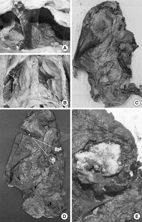
Fig. 3 Dissection of right thoracic cavity. (A) Pleural adhesion (indicated by asterisk). (B) Thoracic cavity after resection of anterior thoracic wall. (C) Right lung (removed). (D) and (E) Exposure of calcified nodules in right lung. (D) Two large calcified nodules (indicated by asterisk; sized over 1 cm in diameter) around pulmonary hilum. (E) Magnified image of calcified nodule (sectioned) in superior segment of right lower lobe.
Reference
-
1. Masson M, Molnár E, Donoghue HD, Besra GS, Minnikin DE, Wu HH, Lee OY, Bull ID, Pálfi G. Osteological and biomolecular evidence of a 7000-year-old case of hypertrophic pulmonary osteopathy secondary to tuberculosis from neolithic hungary. PLoS One. 2013; 8:e78252.2. Bos KI, Harkins KM, Herbig A, Coscolla M, Weber N, Comas I, Forrest SA, Bryant JM, Harris SR, Schuenemann VJ, et al. Pre-Columbian mycobacterial genomes reveal seals as a source of New World human tuberculosis. Nature. 2014; 514:494–497.3. Müller R, Roberts CA, Brown TA. Genotyping of ancient Mycobacterium tuberculosis strains reveals historic genetic diversity. Proc Biol Sci. 2014; 281:20133236.4. Müller R, Roberts CA, Brown TA. Biomolecular identification of ancient Mycobacterium tuberculosis complex DNA in human remains from Britain and continental Europe. Am J Phys Anthropol. 2014; 153:178–189.5. Krogman WM, İşcan MY. The human skeleton in forensic medicine. 1986. Springfield, IL: Charles C Thomas Publisher, LTD.6. Lovejoy CO, Meindl RS, Pryzbeck TR, Mensforth RP. Chronological metamorphosis of the auricular surface of the ilium: a new method for the determination of adult skeletal age at death. Am J Phys Anthropol. 1985; 68:15–28.7. Lamendin H, Baccino E, Humbert JF, Tavernier JC, Nossintchouk RM, Zerilli A. A simple technique for age estimation in adult corpses: the two criteria dental method. J Forensic Sci. 1992; 37:1373.8. Gurney JW, Conces DJ. Pulmonary histoplasmosis. Radiology. 1996; 199:297–306.9. Khan AN, Al-Jahdali HH, Allen CM, Irion KL, Al Ghanem S, Koteyar SS. The calcified lung nodule: What does it mean? Ann Thorac Med. 2010; 5:67.10. Brown K, Mund DF, Aberle DR, Batra P, Young DA. Intrathoracic calcifications: radiographic features and differential diagnoses. Radiographics. 1994; 14:1247–1261.11. Urban BA, Fishman EK, Goldman SM, Scott WW Jr, Jones B, Humphrey RL, Hruban RH. CT evaluation of amyloidosis: spectrum of disease. Radiographics. 1993; 13:1295–1308.12. Palmer PE. Pulmonary tuberculosis--usual and unusual radiographic presentations. Semin Roentgenol. 1979; 14:204–243.13. Lee KS, Im JG. CT in adults with tuberculosis of the chest: characteristic findings and role in management. AJR Am J Roentgenol. 1995; 164:1361–1367.14. Bartels P. Tuberkulose (Wirbelkaries) in der jüngeren Steinzeit 1907 (cited by Formicola V et al. Am J Phys Anthropol. 1987; 72:1–6.15. Formicola V, Milanesi Q, Scarsini C. Evidence of spinal tuberculosis at the beginning of the fourth millennium BC from Arene Candide cave (Liguria, Italy). Am J Phys Anthropol. 1987; 72:1–6.16. Canci A, Minozzi S, Tarli SM. New evidence of tuberculous spondylitis from Neolithic Liguria (Italy). Int J Osteoarchaeol. 1996; 6:497–501.17. Roberts CA, Manchester K. The archaeology of disease. 2007. Ithaca, NY: Cornell University Press.18. Ortner DJ. Identification of pathological conditions in human skeletal remains. 2003. 2nd ed. San Diego, CA: Academic Press.19. Donoghue HD, Spigelman M, Greenblatt CL, Lev-Maor G, Bar-Gal GK, Matheson C, Vernon K, Nerlich AG, Zink AR. Tuberculosis: from prehistory to Robert Koch, as revealed by ancient DNA. Lancet Infect Dis. 2004; 4:584–592.20. Suzuki T, Fujita H, Choi JG. Brief communication: new evidence of tuberculosis from prehistoric Korea-Population movement and early evidence of tuberculosis in far East Asia. Am J Phys Anthropol. 2008; 136:357–360.
- Full Text Links
-
- Actions
-
Cited
- CITED
-
- Close
- Share
-
- Similar articles
-
- Ancient Mitochondrial DNA Analyses of Ascaris Eggs Discovered in Coprolites from Joseon Tomb
- Benign solitary pulmonary nodule: Value of high-resolution CT
- An Unusual Radiologic Manifestation of Pulmonary Tuberculosis with Bilateral Multiple Lung Nodules and Diffuse Alveolar Hemorrhage: A Case Report
- Revealing Joseon period People’s single nucleotide polymorphism associated with lactase gene by ancient DNA analysis of human remains from archaeological sites in Korea
- A Case of Benign Multiple Pulmonary Nodules in a Patient with Osteosarcoma
