J Korean Med Sci.
2015 Dec;30(12):1902-1910. 10.3346/jkms.2015.30.12.1902.
Changes in the Expressions of Iba1 and Calcitonin Gene-Related Peptide in Adjacent Lumbar Spinal Segments after Lumbar Disc Herniation in a Rat Model
- Affiliations
-
- 1Department of Physical Medicine and Rehabilitation, College of Medicine, Daegu Catholic University, Daegu, Korea.
- 2Department of Physical Medicine and Rehabilitation, College of Medicine, Yeungnam University, Daegu, Korea. tra5705@hanmail.net
- 3Medical Devices Clinical Trial Center, Yeungnam University, Daegu, Korea.
- 4Department of Anatomy and Cell Biology, School of Medicine, Hanyang University, Seoul, Korea.
- KMID: 2359978
- DOI: http://doi.org/10.3346/jkms.2015.30.12.1902
Abstract
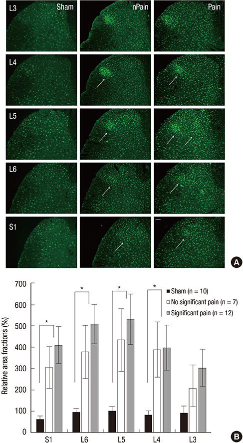
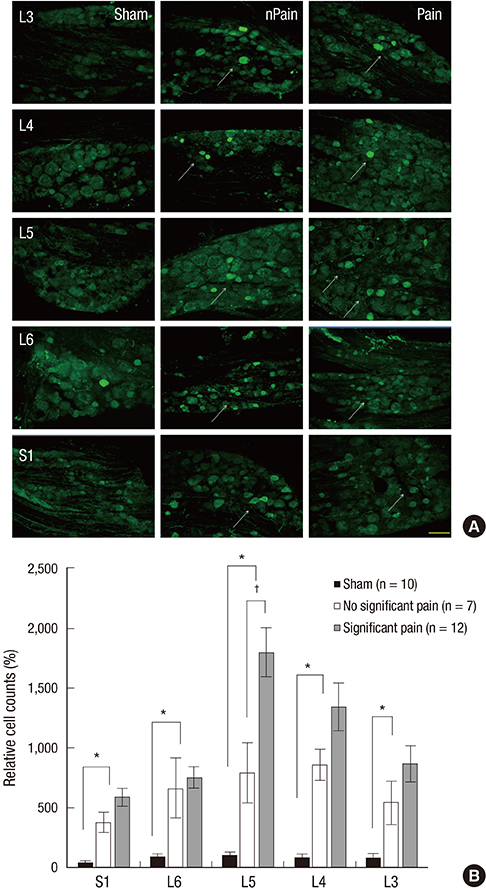
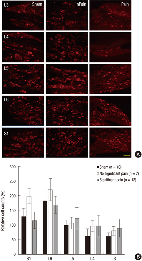
- Lumbar disc herniation is commonly encountered in clinical practice and can induce sciatica due to mechanical and/or chemical irritation and the release of proinflammatory cytokines. However, symptoms are not confined to the affected spinal cord segment. The purpose of this study was to determine whether multisegmental molecular changes exist between adjacent lumbar spinal segments using a rat model of lumbar disc herniation. Twenty-nine male Sprague-Dawley rats were randomly assigned to either a sham-operated group (n=10) or a nucleus pulposus (NP)-exposed group (n=19). Rats in the NP-exposed group were further subdivided into a significant pain subgroup (n=12) and a no significant pain subgroup (n=7) using mechanical pain thresholds determined von Frey filaments. Immunohistochemical stainings of microglia (ionized calcium-binding adapter molecule 1; Iba1), astrocytes (glial fibrillary acidic protein; GFAP), calcitonin gene-related peptide (CGRP), and transient receptor potential vanilloid 1 (TRPV1) was performed in spinal dorsal horns and dorsal root ganglions (DRGs) at 10 days after surgery. It was found immunoreactivity for Iba1-positive microglia was higher in the L5 (P=0.004) dorsal horn and in the ipsilateral L4 (P=0.009), L6 (P=0.002), and S1 (P=0.002) dorsal horns in the NP-exposed group than in the sham-operated group. The expression of CGRP was also significantly higher in ipsilateral L3, L4, L6, and S1 segments and in L5 DRGs at 10 days after surgery in the NP-exposed group than in the sham-operated group (P<0.001). Our results indicate that lumbar disc herniation upregulates microglial activity and CGRP expression in many adjacent and ipsilateral lumbar spinal segments.
Keyword
MeSH Terms
-
Animals
Astrocytes/metabolism
Calcitonin Gene-Related Peptide/*metabolism
Calcium-Binding Proteins/*metabolism
Disease Models, Animal
Ganglia, Spinal/metabolism
Humans
Immunohistochemistry
Intervertebral Disc Displacement/*metabolism
Lumbar Vertebrae/*metabolism
Male
Microfilament Proteins/*metabolism
Microglia/metabolism
Neuralgia/metabolism
Rats
Rats, Sprague-Dawley
Spinal Cord Dorsal Horn/metabolism
Up-Regulation
Calcitonin Gene-Related Peptide
Calcium-Binding Proteins
Microfilament Proteins
Figure
Cited by 1 articles
-
Toll-like receptor 4/nuclear factor-kappa B pathway is involved in radicular pain by encouraging spinal microglia activation and inflammatory response in a rat model of lumbar disc herniation
Lirong Zhu, Yangliang Huang, Yuming Hu, Qian Tang, Yi Zhong
Korean J Pain. 2021;34(1):47-57. doi: 10.3344/kjp.2021.34.1.47.
Reference
-
1. Olmarker K, Brisby H, Yabuki S, Nordborg C, Rydevik B. The effects of normal, frozen, and hyaluronidase-digested nucleus pulposus on nerve root structure and function. Spine (Phila Pa 1976). 1997; 22:471–475.2. Olmarker K, Larsson K. Tumor necrosis factor alpha and nucleus-pulposus-induced nerve root injury. Spine (Phila Pa 1976). 1998; 23:2538–2544.3. Olmarker K, Rydevik B, Nordborg C. Autologous nucleus pulposus induces neurophysiologic and histologic changes in porcine cauda equina nerve roots. Spine (Phila Pa 1976). 1993; 18:1425–1432.4. Igarashi T, Kikuchi S, Shubayev V, Myers RR. 2000 Volvo Award winner in basic science studies: exogenous tumor necrosis factor-alpha mimics nucleus pulposus-induced neuropathology Molecular, histologic, and behavioral comparisons in rats. Spine (Phila Pa 1976). 2000; 25:2975–2980.5. Kallakuri S, Takebayashi T, Ozaktay AC, Chen C, Yang S, Wooley PH, Cavanaugh JM. The effects of epidural application of allografted nucleus pulposus in rats on cytokine expression, limb withdrawal and nerve root discharge. Eur Spine J. 2005; 14:956–964.6. Ohtori S, Inoue G, Koshi T, Ito T, Doya H, Saito T, Moriya H, Takahashi K. Up-regulation of acid-sensing ion channel 3 in dorsal root ganglion neurons following application of nucleus pulposus on nerve root in rats. Spine (Phila Pa 1976). 2006; 31:2048–2052.7. Ito T, Ohtori S, Inoue G, Koshi T, Doya H, Ozawa T, Saito T, Moriya H, Takahashi K. Glial phosphorylated p38 MAP kinase mediates pain in a rat model of lumbar disc herniation and induces motor dysfunction in a rat model of lumbar spinal canal stenosis. Spine (Phila Pa 1976). 2007; 32:159–167.8. Badalamente MA, Dee R, Ghillani R, Chien PF, Daniels K. Mechanical stimulation of dorsal root ganglia induces increased production of substance P: a mechanism for pain following nerve root compromise? Spine (Phila Pa 1976). 1987; 12:552–555.9. Kobayashi S, Uchida K, Kokubo Y, Takeno K, Yayama T, Miyazaki T, Nakajima H, Nomura E, Mwaka E, Baba H. Synapse involvement of the dorsal horn in experimental lumbar nerve root compression: a light and electron microscopic study. Spine (Phila Pa 1976). 2008; 33:716–723.10. Kim SJ, Kim WR, Kim HS, Park HW, Cho YW, Jang SH, Hwang SJ, Ahn SH. Abnormal spontaneous activities on needle electromyography and their relation with pain behavior and nerve fiber pathology in a rat model of lumbar disc herniation. Spine (Phila Pa 1976). 2011; 36:E1562–E1567.11. Park HW, Ahn SH, Kim SJ, Seo JM, Cho YW, Jang SH, Hwang SJ, Kwak SY. Changes in spinal cord expression of fractalkine and its receptor in a rat model of disc herniation by autologous nucleus pulposus. Spine (Phila Pa 1976). 2011; 36:E753–E760.12. Guo J, Fu X, Cui X, Fan M. Contributions of purinergic P2X3 receptors within the midbrain periaqueductal gray to diabetes-induced neuropathic pain. J Physiol Sci. 2015; 65:99–104.13. Benoist M. The natural history of lumbar disc herniation and radiculopathy. Joint Bone Spine. 2002; 69:155–160.14. Ahn SH, Cho YW, Ahn MW, Jang SH, Sohn YK, Kim HS. mRNA expression of cytokines and chemokines in herniated lumbar intervertebral discs. Spine (Phila Pa 1976). 2002; 27:911–917.15. Shubayev VI, Myers RR. Anterograde TNF alpha transport from rat dorsal root ganglion to spinal cord and injured sciatic nerve. Neurosci Lett. 2002; 320:99–101.16. Tsuda M, Inoue K, Salter MW. Neuropathic pain and spinal microglia: a big problem from molecules in "small" glia. Trends Neurosci. 2005; 28:101–107.17. Watkins LR, Maier SF. Glia: a novel drug discovery target for clinical pain. Nat Rev Drug Discov. 2003; 2:973–985.18. Ohtori S, Takahashi K, Aoki Y, Doya H, Ozawa T, Saito T, Moriya H. Spinal neural cyclooxygenase-2 mediates pain caused in a rat model of lumbar disk herniation. J Pain. 2004; 5:385–391.19. Ding M, Hart RP, Jonakait GM. Tumor necrosis factor-alpha induces substance P in sympathetic ganglia through sequential induction of interleukin-1 and leukemia inhibitory factor. J Neurobiol. 1995; 28:445–454.20. Hua XY, Chen P, Fox A, Myers RR. Involvement of cytokines in lipopolysaccharide-induced facilitation of CGRP release from capsaicin-sensitive nerves in the trachea: studies with interleukin-1beta and tumor necrosis factor-alpha. J Neurosci. 1996; 16:4742–4748.21. Fukuoka T, Kondo E, Dai Y, Hashimoto N, Noguchi K. Brain-derived neurotrophic factor increases in the uninjured dorsal root ganglion neurons in selective spinal nerve ligation model. J Neurosci. 2001; 21:4891–4900.22. Li H, Li Q, Xie K, Feng S, Wang P, Ma X. Expression of c-Fos and c-Jun in adjacent cervical spinal cord segments following C nerve root rhizotomy in rats: Indication of a neural pathway between adjacent cervical spinal cord segments. Exp Ther Med. 2013; 6:373–377.23. Colangelo AM, Bianco MR, Vitagliano L, Cavaliere C, Cirillo G, De Gioia L, Diana D, Colombo D, Redaelli C, Zaccaro L, et al. A new nerve growth factor-mimetic peptide active on neuropathic pain in rats. J Neurosci. 2008; 28:2698–2709.24. Cirillo G, Cavaliere C, Bianco MR, De Simone A, Colangelo AM, Sellitti S, Alberghina L, Papa M. Intrathecal NGF administration reduces reactive astrocytosis and changes neurotrophin receptors expression pattern in a rat model of neuropathic pain. Cell Mol Neurobiol. 2010; 30:51–62.25. Cirillo G, Bianco MR, Colangelo AM, Cavaliere C, Daniele de L, Zaccaro L, Alberghina L, Papa M. Reactive astrocytosis-induced perturbation of synaptic homeostasis is restored by nerve growth factor. Neurobiol Dis. 2011; 41:630–639.26. Giaume C, Koulakoff A, Roux L, Holcman D, Rouach N. Astroglial networks: a step further in neuroglial and gliovascular interactions. Nat Rev Neurosci. 2010; 11:87–99.27. Todd AJ. Neuronal circuitry for pain processing in the dorsal horn. Nat Rev Neurosci. 2010; 11:823–836.28. Meisner JG, Marsh AD, Marsh DR. Loss of GABAergic interneurons in laminae I-III of the spinal cord dorsal horn contributes to reduced GABAergic tone and neuropathic pain after spinal cord injury. J Neurotrauma. 2010; 27:729–737.29. Cirillo G, Colangelo AM, Bianco MR, Cavaliere C, Zaccaro L, Sarmientos P, Alberghina L, Papa M. BB14, a Nerve Growth Factor (NGF)-like peptide shown to be effective in reducing reactive astrogliosis and restoring synaptic homeostasis in a rat model of peripheral nerve injury. Biotechnol Adv. 2012; 30:223–232.30. Hatashita S, Sekiguchi M, Kobayashi H, Konno S, Kikuchi S. Contralateral neuropathic pain and neuropathology in dorsal root ganglion and spinal cord following hemilateral nerve injury in rats. Spine (Phila Pa 1976). 2008; 33:1344–1351.31. Kim DS, Figueroa KW, Li KW, Boroujerdi A, Yolo T, Luo ZD. Profiling of dynamically changed gene expression in dorsal root ganglia post peripheral nerve injury and a critical role of injury-induced glial fibrillary acidic protein in maintenance of pain behaviors [corrected]. Pain. 2009; 143:114–122.32. Li Y, Xi C, Niu M, Liu X, Chi Z, Wang X, Yan J. Contralateral neuropathology in dorsal root ganglia in a rat model of noncompressive disc herniation. Neurosci Lett. 2011; 493:49–54.33. Rothman SM, Guarino BB, Winkelstein BA. Spinal microglial proliferation is evident in a rat model of painful disc herniation both in the presence of behavioral hypersensitivity and following minocycline treatment sufficient to attenuate allodynia. J Neurosci Res. 2009; 87:2709–2717.34. Echeverry S, Shi XQ, Zhang J. Characterization of cell proliferation in rat spinal cord following peripheral nerve injury and the relationship with neuropathic pain. Pain. 2008; 135:37–47.35. Cho HK, Cho YW, Kim EH, Sluijter ME, Hwang SJ, Ahn SH. Changes in pain behavior and glial activation in the spinal dorsal horn after pulsed radiofrequency current administration to the dorsal root ganglion in a rat model of lumbar disc herniation: laboratory investigation. J Neurosurg Spine. 2013; 19:256–263.36. Matta JA, Ahern GP. TRPV1 and synaptic transmission. Curr Pharm Biotechnol. 2011; 12:95–101.37. Tominaga M, Caterina MJ, Malmberg AB, Rosen TA, Gilbert H, Skinner K, Raumann BE, Basbaum AI, Julius D. The cloned capsaicin receptor integrates multiple pain-producing stimuli. Neuron. 1998; 21:531–543.38. Michael GJ, Priestley JV. Differential expression of the mRNA for the vanilloid receptor subtype 1 in cells of the adult rat dorsal root and nodose ganglia and its downregulation by axotomy. J Neurosci. 1999; 19:1844–1854.39. Chen Y, Willcockson HH, Valtschanoff JG. Influence of the vanilloid receptor TRPV1 on the activation of spinal cord glia in mouse models of pain. Exp Neurol. 2009; 220:383–390.40. Patwardhan AM, Scotland PE, Akopian AN, Hargreaves KM. Activation of TRPV1 in the spinal cord by oxidized linoleic acid metabolites contributes to inflammatory hyperalgesia. Proc Natl Acad Sci U S A. 2009; 106:18820–18824.
- Full Text Links
- Actions
-
Cited
- CITED
-
- Close
- Share
- Similar articles
-
- The Effect of GCSB-5 a New Herbal Medicine on Changes in Pain Behavior and Neuroglial Activation in a Rat Model of Lumbar Disc Herniation
- Effect of Antiandrogen on Calcitonin Gene-related Peptide mRNA Expression ofthe Rat Cremaster Nucleus
- High Lumbar Disc Herniation Treated With an Anterolateral Approach: Case Report
- High Lumbar Disc Herniation Treated with A Modified Posterolateral Approach: Case Report
- Analysis of MRI Findings of Adolescent Lumbar Disc Herniation (Comparision with Adult Lumbar Disc Herniation Findings)

