Neural Axis Metastasis from Metachronous Pulmonary Basaloid Carcinoma Developed after Chemotherapy & Radiation Therapy of Uterine Cervical Carcinoma
- Affiliations
-
- 1Department of Neurosurgery, Soonchunhyang University College of Medicine, Bucheon Hospital, Bucheon, Korea. neuri71@schmc.ac.kr
- 2Department of Pathology, Soonchunhyang University College of Medicine, Bucheon Hospital, Bucheon, Korea.
- 3Department of Obstetrics and Gynecology, Soonchunhyang University College of Medicine, Bucheon Hospital, Bucheon, Korea.
- KMID: 2356795
- DOI: http://doi.org/10.13004/kjnt.2016.12.2.167
Abstract
- Multiple primary or secondary malignancies after anticancer therapy were recently reported to be increasing in frequency. The authors describe a case of metachronous metastatic pulmonary basaloid carcinoma to the central nervous system that was discovered after chemotherapy and radiation therapy for cervical uterine carcinoma. Two different types of cancer developed within some interval. There's the possibility that a secondary pulmonary neoplasm developed after the chemotherapy and radiotherapy conducted as cervical cancer treatment.
MeSH Terms
Figure
-
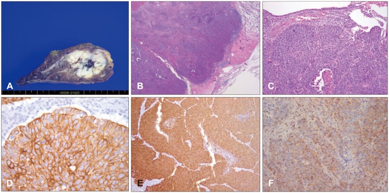
FIGURE 1 Gross findings of wedge-resected lung show a 2.8×2.7-cm, well-circumscribed, whitish, firm, lobulated mass with hemorrhage and necrosis. (B) Microscopic findings show a well-circumscribed, lobulated, solid tumor with peripheral palisading and edematous lung parenchyma (hematoxylin and eosin [H & E] stain, ×40). (C) The tumor shows small cuboidal-to-fusiform tumor cells with moderately hyperchromatic nuclei and nuclear pleomorphism as well as multifocal necrosis (H & E stain, ×100). (D) Immunohistochemistry (IHC) of cytokeratin 7 reveals diffuse positivity in tumor cells (cytokeratin 7, ×200). (E) Immunohistochemistry of P16 reveals diffuse positivity in tumor cells (P16, ×100). (F) IHC of CD99 reveals dot-like positivity in tumor cell cytoplasm (CD99, ×200).
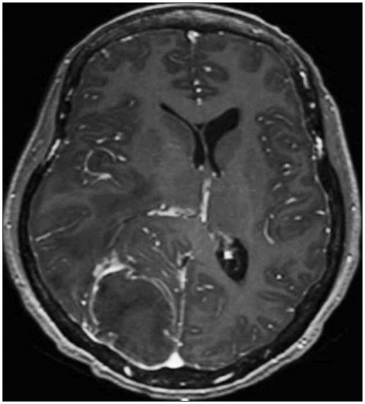
FIGURE 2 Brain magnetic resonance imaging with contrast enhancement. Shows 6×4-cm rim enhanced mass with vasogenic edema in the right parieto-occipital lobe.
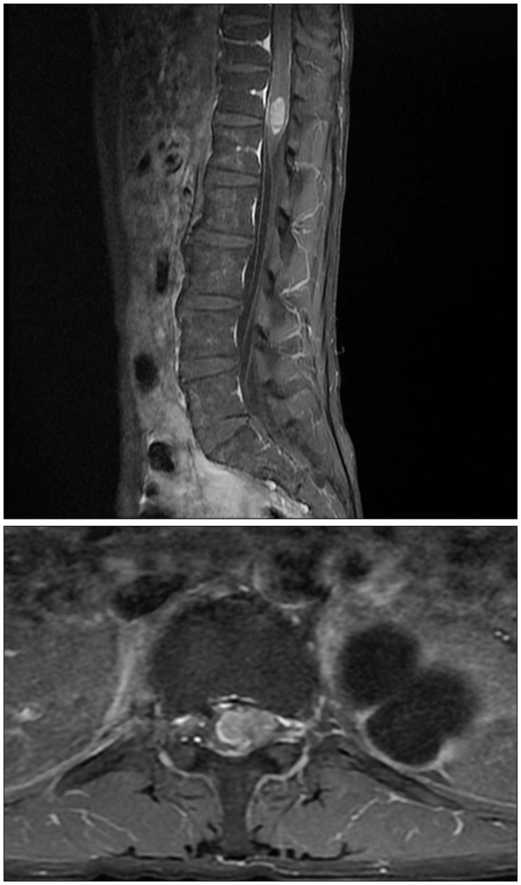
FIGURE 3 Lumbar spine magnetic resonance imaging with contrast enhancement. Well enhancing contour bulging mass in the conus medullaris of the spinal cord at the T12-L1 level.
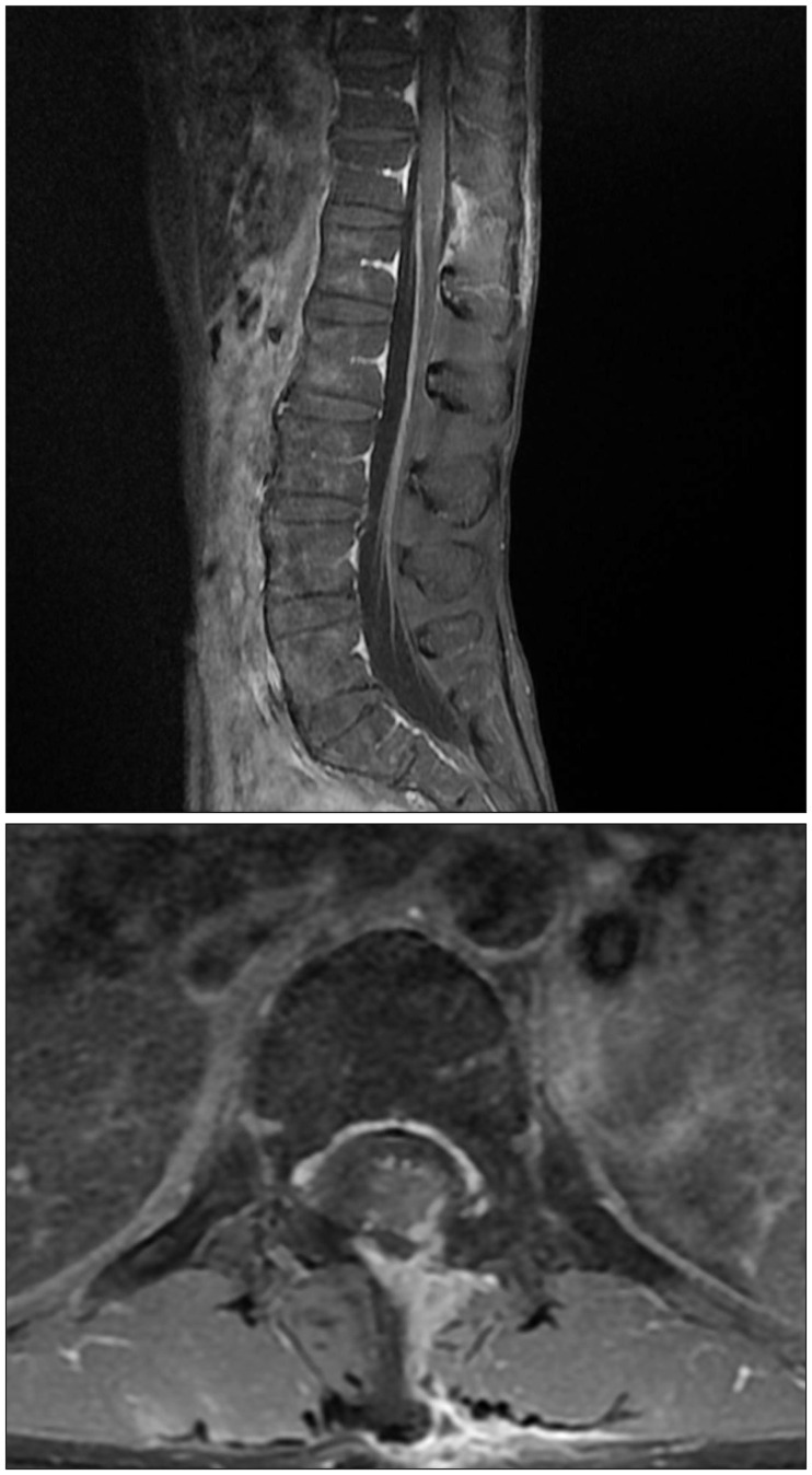
FIGURE 4 Lumbar-spine magnetic resonance imaging with contrast enhancement, postoperative status. Near total removal of metastatic lesion of the spinal cord.
Reference
-
1. Alexander MJ, DeSalles AA, Tomiyasu U. Multiple radiation-induced intracranial lesions after treatment for pituitary adenoma. Case report. J Neurosurg. 1998; 88:111–115. PMID: 9420081.2. Amirjamshidi A, Abbassioun K. Radiation-induced tumors of the central nervous system occurring in childhood and adolescence. Four unusual lesions in three patients and a review of the literature. Childs Nerv Syst. 2000; 16:390–397. PMID: 10958546.3. Argani P, Laé M, Ballard ET, Amin M, Manivel C, Hutchinson B, et al. Translocation carcinomas of the kidney after chemotherapy in childhood. J Clin Oncol. 2006; 24:1529–1534. PMID: 16575003.
Article4. Bhagavathi S, Chang CH. Multicentric basaloid carcinoma of lung clinically mimicking metastatic carcinoma: a case report. Int J Surg Pathol. 2009; 17:68–71. PMID: 18480394.
Article5. Brambilla E, Moro D, Veale D, Brichon PY, Stoebner P, Paramelle B, et al. Basal cell (basaloid) carcinoma of the lung: a new morphologic and phenotypic entity with separate prognostic significance. Hum Pathol. 1992; 23:993–1003. PMID: 1381335.
Article6. Demandante CG, Troyer DA, Miles TP. Multiple primary malignant neoplasms: case report and a comprehensive review of the literature. Am J Clin Oncol. 2003; 26:79–83. PMID: 12576929.7. Findlay JM, Bernstein M, Vanderlinden RG, Resch L. Microsurgical resection of solitary intramedullary spinal cord metastases. Neurosurgery. 1987; 21:911–915. PMID: 3437960.
Article8. Grem JL, Burgess J, Trump DL. Clinical features and natural history of intramedullary spinal cord metastasis. Cancer. 1985; 56:2305–2314. PMID: 4052974.
Article9. Hargraves RW, Cotelingam JD, Harris RD, Pulliam MW. Unusual metastasis to the cauda equina: case report. Neurosurgery. 1986; 19:828–830. PMID: 3785635.
Article10. Hope AJ, Mansur DB, Tu PH, Simpson JR. Metachronous secondary atypical meningioma and anaplastic astrocytoma after postoperative craniospinal irradiation for medulloblastoma. Childs Nerv Syst. 2006; 22:1201–1207. PMID: 16570196.
Article11. Okamoto H, Shinkai T, Matsuno Y, Saijo N. Intradural parenchymal involvement in the spinal subarachnoid space associated with primary lung cancer. Cancer. 1993; 72:2583–2588. PMID: 8402479.
Article12. Sasayama T, Nishihara M, Tanaka K, Mizukawa K, Ehara K, Kanomata N, et al. Two metachronous tumors induced by radiation therapy: case report and review of the literature. J Neurooncol. 2008; 88:315–320. PMID: 18373066.
Article
- Full Text Links
-
- Actions
-
Cited
- CITED
-
- Close
- Share
-
- Similar articles
-
- A Case of Postirradiation Uterine Papillary Serous Carcinoma
- Solitary Cerebellar Metastasis from Primary Uterine Cervical Carcinoma: A Case Report
- Basaloid-Squamous Carcinoma of the Esophagus: A case report
- Pure Basaloid Squamous Cell Carcinoma of the Uterine Cervix: A Case Report
- A case of ovarian metastasis of the recurrent uterine cervical cancer after adjuvant radiation therapy
