Shunt Overdrainage Caused by Displacement of the Pressure Control Cam after Pressure Adjustment
- Affiliations
-
- 1Department of Neurosurgery, Daegu Fatima Hospital, Daegu, Korea. paulyoonsoolee@hanmail.net
- KMID: 2356794
- DOI: http://doi.org/10.13004/kjnt.2016.12.2.163
Abstract
- Although the Codman-Hakim programmable valve is one of most popular shunt systems used in the clinical practice for the treatment of hydrocephalus, malfunctions related with this system have been also reported which lead to underdrainage or overdrainage of the cerebrospinal fluid. While obstruction of the ventricular catheter by tissue materials or hematoma and catheter disconnection are relatively common, the malfunction of the valve itself is rare. Herein, we report on a rare case of shunt overdrainage caused by displacement of the pressure control cam after pressure adjustment. A 57-year-old female, who underwent a ventriculoperitoneal shunt eight years ago, experienced aggravating symptoms of shunt overdrainage after pressure adjustment. Displacement of the pressure control cam was revealed on the X-ray, and a shunt revision was performed. The purpose of this report is to provide a working knowledge of the valve structure and to enhance the ability to interpret the valve setting on an X-ray for diagnosis of valve malfunction.
MeSH Terms
Figure
-
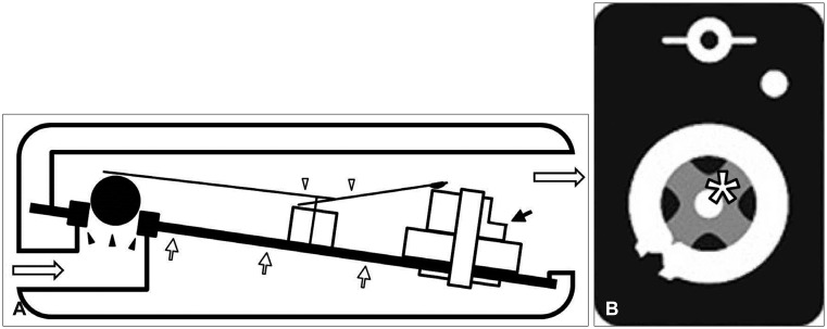
FIGURE 1 (A) Illustration depicts the structure of the Codman-Hakim programmable valve from the side view. The flat spring (white arrowheads) presses the valve ball landed on a matching valve seat (black arrowheads) through which cerebrospinal fluid (CSF) is passed distally as well as the pressure control cam (short black arrow) which controls the intensity of the flat spring. These structures are built on the base plate (short white arrows). Long white arrows indicate the direction of CSF flow. (B) Illustration depicts the valve from the top view. The bottom side of the cam has a protrusion of "X" (white asterisk) in the center and this part is fixed to a corresponding X-shaped slit on the base plate by the pressure of the flat spring.
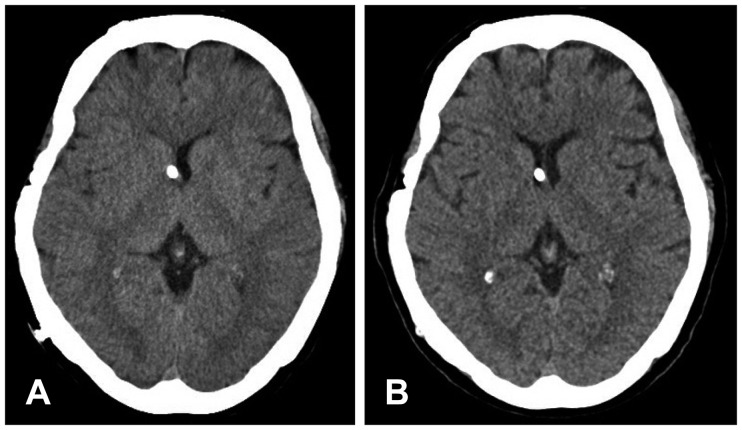
FIGURE 2 (A) A brain computed tomography (CT) before shunt revision shows a slit-ventricle. (B) A brain CT after shunt revision shows recovery from the slit-ventricle.
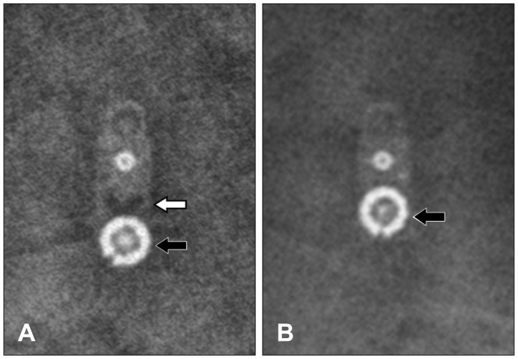
FIGURE 3 (A) A skull X-ray shows that the pressure control cam (black arrow) is detached from the base plate and displaced distally. Note the empty X-shaped slit (white arrow) on the base plate. (B) Another skull X-ray of a normal valve with pressure setting at 10 mmH2O is also provided for comparison. Note the correct location of the cam (black arrow) on the X-ray.
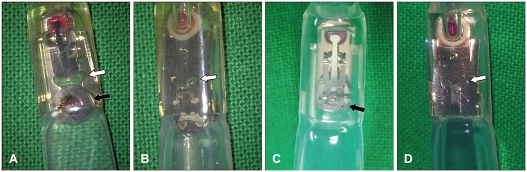
FIGURE 4 Intraoperative photographs reveal the detachment of the cam (black arrow) from the base plate leaving the X-shaped slit (white arrow) on the base plate empty. (A) Top view. (B) Bottom view. Magnified photographs of a normal valve are also provided for comparison. (C) Top view. (D) Bottom view. Note the correct location of the cam (black arrow) on the base plate. The protrusion of "X" is properly landed on the X-shaped slit (white arrow).
Reference
-
1. Black PM, Hakim R, Bailey NO. The use of the Codman-Medos Programmable Hakim valve in the management of patients with hydrocephalus: illustrative cases. Neurosurgery. 1994; 34:1110–1113. PMID: 8084404.2. Kurosaki K, Hamada H, Hayashi N, Kurimoto M, Hirashima Y, Endo S. A rare case of shunt malfunction attributable to blockage of a Codman-Hakim programmable shunt valve. Childs Nerv Syst. 2002; 18:183–185. PMID: 11981632.
Article3. Lollis SS, Mamourian AC, Vaccaro TJ, Duhaime AC. Programmable CSF shunt valves: radiographic identification and interpretation. AJNR Am J Neuroradiol. 2010; 31:1343–1346. PMID: 20150313.
Article4. Okazaki T, Oki S, Migita K, Kurisu K. A rare case of shunt malfunction attributable to a broken Codman-Hakim programmable shunt valve after a blow to the head. Pediatr Neurosurg. 2005; 41:241–243. PMID: 16195675.
Article5. Shellock FG, Wilson SF, Mauge CP. Magnetically programmable shunt valve: MRI at 3-Tesla. Magn Reson Imaging. 2007; 25:1116–1121. PMID: 17707175.
Article
- Full Text Links
-
- Actions
-
Cited
- CITED
-
- Close
- Share
-
- Similar articles
-
- Clinical Experience with the Programmable Valve for Hydrocephalus Patients
- Efficacy of the Programmable Valve in the Treatment of Hydrocephalus
- Acute Epidural Hematoma Following Ventriculo-Peritoneal(V-P) shunt Operation
- Cervical Myelopathy Due to Epidural Hematoma at the Cervicomedullary Junction Associated With Ventriculoperitoneal Shunt Overdrainage: A Case Report
- Cervical Myelopathy Caused by a Ventriculoperitoneal Shunt: A Case Report
