J Korean Soc Radiol.
2016 Nov;75(5):404-409. 10.3348/jksr.2016.75.5.404.
Magnetic Resonance Imaging Findings of Solitary Infantile Myofibromatosis of the Skull: A Case Report
- Affiliations
-
- 1Department of Radiology, College of Medicine, Yeungnam University, Daegu, Korea. khcho@med.yu.ac.kr
- 2Department of Pathology, College of Medicine, Yeungnam University, Daegu, Korea.
- KMID: 2355996
- DOI: http://doi.org/10.3348/jksr.2016.75.5.404
Abstract
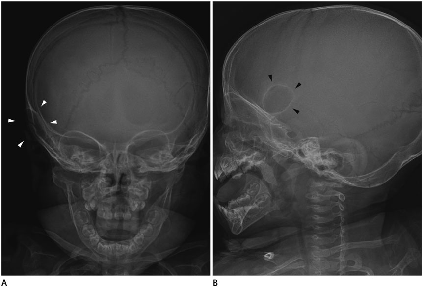
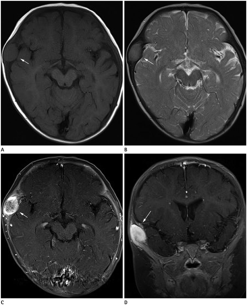
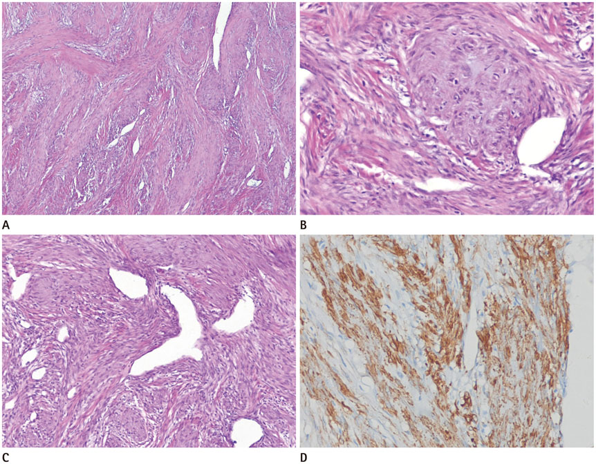
- Infantile myofibromatosis is a rare, benign mesenchymal disorder of early childhood characterized by solitary or multiple benign myofibroblastic tumors. The tumors may involve the skin, subcutaneous tissue, muscle, bone and visceral organs. We report magnetic resonance imaging findings of solitary infantile myofibromatosis arising in the temporal bone of a ten-month-old boy, and the diagnosis was confirmed by surgical excision and histopathological examination.
MeSH Terms
Figure
Reference
-
1. Inwards CY, Unni KK, Beabout JW, Shives TC. Solitary congenital fibromatosis (infantile myofibromatosis) of bone. Am J Surg Pathol. 1991; 15:935–941.2. Chung EB, Enzinger FM. Infantile myofibromatosis. Cancer. 1981; 48:1807–1818.3. Murphey MD, Ruble CM, Tyszko SM, Zbojniewicz AM, Potter BK, Miettinen M. From the archives of the AFIP: musculoskeletal fibromatoses: radiologic-pathologic correlation. Radiographics. 2009; 29:2143–2173.4. Okamoto K, Ito J, Takahashi H, Emura I, Mori H, Furusawa T, et al. Solitary myofibromatosis of the skull. Eur Radiol. 2000; 10:170–174.5. Tsuji M, Inagaki T, Kasai H, Yamanouchi Y, Kawamoto K, Uemura Y. Solitary myofibromatosis of the skull: a case report and review of literature. Childs Nerv Syst. 2004; 20:366–369.6. Merciadri P, Pavanello M, Nozza P, Consales A, Ravegnani GM, Piatelli G, et al. Solitary infantile myofibromatosis of the cranial vault: case report. Childs Nerv Syst. 2011; 27:643–647.7. Koujok K, Ruiz RE, Hernandez RJ. Myofibromatosis: imaging characteristics. Pediatr Radiol. 2005; 35:374–380.8. Morón FE, Morriss MC, Jones JJ, Hunter JV. Lumps and bumps on the head in children: use of CT and MR imaging in solving the clinical diagnostic dilemma. Radiographics. 2004; 24:1655–1674.9. Yim Y, Moon WJ, An HS, Cho J, Rho MH. Imaging findings of various calvarial bone lesions with a focus on osteolytic lesions. J Korean Soc Radiol. 2016; 74:43–54.10. D'Ambrosio N, Lyo JK, Young RJ, Haque SS, Karimi S. Imaging of metastatic CNS neuroblastoma. AJR Am J Roentgenol. 2010; 194:1223–1229.


