J Cerebrovasc Endovasc Neurosurg.
2016 Jun;18(2):115-119. 10.7461/jcen.2016.18.2.115.
Neurological Deterioration after Decompressive Suboccipital Craniectomy in a Patient with a Brainstem-compressing Thrombosed Giant Aneurysm of the Vertebral Artery
- Affiliations
-
- 1Department of Neurosurgery, Gangnam Severance Hospital, Yonsei University College of Medicine, Seoul, Korea. ns.joonho.chung@gmail.com
- 2Department of Neurosurgery, Dongsan Medical Center, College of Medicine, Keimyung University, Daegu, Korea.
- 3Severance Institute for Vascular and Metabolic Research, Yonsei University College of Medicine, Seoul, Korea.
- KMID: 2354886
- DOI: http://doi.org/10.7461/jcen.2016.18.2.115
Abstract
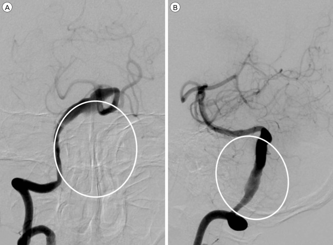
- We experienced a case of neurological deterioration after decompressive suboccipital craniectomy (DSC) in a patient with a brainstem-compressing thrombosed giant aneurysm of the vertebral artery (VA). A 60-year-old male harboring a thrombosed giant aneurysm (about 4 cm) of the right vertebral artery presented with quadriparesis. We treated the aneurysm by endovascular coil trapping of the right VA and expected the aneurysm to shrink slowly. After 7 days, however, he suffered aggravated symptoms as his aneurysm increased in size due to internal thrombosis. The medulla compression was aggravated, and so we performed DSC with C1 laminectomy. After the third post-operative day, unfortunately, his neurologic symptoms were more aggravated than in the pre-DSC state. Despite of conservative treatment, neurological symptoms did not improve, and microsurgical aneurysmectomy was performed for the medulla decompression. Unfortunately, the post-operative recovery was not as good as anticipated. DSC should not be used to release the brainstem when treating a brainstem-compressing thrombosed giant aneurysm of the VA.
MeSH Terms
Figure
Reference
-
1. Chotai S, Kshettry VR, Lamki T, Ammirati M. Surgical outcomes using wide suboccipital decompression for adult Chiari I malformation with and without syringomyelia. Clin Neurol Neurosurg. 2014; 5. 120:129–135. PMID: 24656777.
Article2. Drake CG. Giant intracranial aneurysms: experience with surgical treatment in 174 patients. Clin Neurosurg. 1979; 26:12–95. PMID: 544122.
Article3. Ganti SR, Steinberger A, McMurtry JG 3rd, Hilal SK. Computed tomographic demonstration of giant aneurysms of the vertebrobasilar system: report of eight cases. Neurosurgery. 1981; 9. 9(3):261–267. PMID: 7301068.4. Hecht ST, Horton JA, Yonas H. Growth of a thrombosed giant vertebral artery aneurysm after parent artery occlusion. AJNR Am J Neuroradiol. 1991; May-Jun. 12(3):449–451. PMID: 2058491.5. Iihara K1, Murao K, Sakai N, Soeda A, Ishibashi-Ueda H, Yutani C, et al. Continued growth of and increased symptoms from a thrombosed giant aneurysm of the vertebral artery after complete endovascular occlusion and trapping: the role of vasa vasorum. Case report. J Neurosurg. 2003; 2. 98(2):407–413. PMID: 12593631.6. Nagahiro S, Takada A, Goto S, Kai Y, Ushio Y. Thrombosed growing giant aneurysms of the vertebral artery: growth mechanism and management. J Neurosurg. 1995; 5. 82(5):796–801. PMID: 7714605.
Article7. Pfefferkorn T, Eppinger U, Linn J, Birnbaum T, Herzog J, Straube A, et al. Long-term outcome after suboccipital decompressive craniectomy for malignant cerebellar infarction. Stroke. 2009; 7. 40(9):3045–3050. PMID: 19574555.
Article8. Ragel BT, Klimo P Jr, Martin JE, Teff RJ, Bakken HE, Armonda RA. Wartime decompressive craniectomy: technique and lessons learned. Neurogurg Focus. 2010; 5. 28(5):E2.
Article9. Ratliff JK, Cooper PR. Cervical laminoplasty: a critical review. J Neurosurg. 2003; 4. 98(3 Suppl):230–238. PMID: 12691377.
Article10. Suda K, Abumi K, Ito M, Shono Y, Kaneda K, Fujiya M. Local kyphosis reduces surgical outcomes of expansive open-door laminoplasty for cervical spondylotic myelophathy. Spine (Phila Pa 1976). 2003; 6. 28(12):1258–1262. PMID: 12811268.11. Sugita K, Kobayashi S, Takemae T, Tanaka Y, Okudera H, Ohsawa M. Giant aneurysms of the vertebral artery. Report of five cases. J Neurosurg. 1988; 6. 68(6):960–966. PMID: 3373290.
- Full Text Links
- Actions
-
Cited
- CITED
-
- Close
- Share
- Similar articles
-
- Unusual Clinical Course of Giant Vertebral Artery Aneurysm after Proximal Artery Embolization: Case Report
- Surgical Management of Acute Infarction
- Surgical Treatment by the Far Lateral Inferial Suboccipital Approach for the Distal Vertebral Artery Aneurysm
- Strategic Dual Approach for the Management of a Symptomatic Giant Partially Thrombosed Aneurysm at the Basilar Tip - Integrating Intrasaccular Flow Diversion and Endovascular Flow Reversal
- A Case Report of Giant Posterior Inferior Cerebellar Artery Aneurysm Simulating a Posterior Fossa Tumor


