J Clin Neurol.
2016 Jul;12(3):289-294. 10.3988/jcn.2016.12.3.289.
Diagnostic Significance of Ultrasonographic Measurements and Median-Ulnar Ratio in Carpal Tunnel Syndrome: Correlation with Nerve Conduction Studies
- Affiliations
-
- 1Department of Physical Medicine and Rehabilitation, Gaziosmanpasa Taksim Education and Research Hospital, Istanbul, Turkey. yurdakul_ozan@yahoo.com
- 2Department of Physical Medicine and Rehabilitation, Haydarpasa Numune Education and Research Hospital, Istanbul, Turkey.
- 3Department of Neurology, Haydarpasa Numune Education and Research Hospital, Istanbul, Turkey.
- KMID: 2354115
- DOI: http://doi.org/10.3988/jcn.2016.12.3.289
Abstract
- BACKGROUND AND PURPOSE
We determined the reliability of ultrasonography (US) measurements for diagnosing carpal tunnel syndrome (CTS) and their correlation with symptom duration and electrophysiology findings. We determined whether the ratio of the median-to-ulnar cross-sectional areas (CSAs) can support CTS diagnoses.
METHODS
The pisiform CSA (CSA(pisiform)), swelling ratio (SR), palmar bowing, and CSA(pisiform)/ulnar CSA (CSA(ulnar)) measurements made in two subgroups of CTS patients (having sensory affection alone or having both sensory and motor affection) were compared with controls. CSA(ulnar) was measured in Guyon's canal at the level of most-protuberant portion of the pisiform bone.
RESULTS
The values of all of the measured US parameters were higher in patients with CTS (n=50) than in controls (n=62). CSA(pisiform) could be used to diagnose CTS of mild severity. All of the parameters were positively correlated with the distal latency of the compound muscle action potential, and all of them except for SR were negatively correlated with the sensory nerve conduction velocity. A CSA(pisiform)/CSA(ulnar) ratio of ≥1.79 had a sensitivity of 70% and a specificity of 76% for diagnosing CTS.
CONCLUSIONS
Only CSA(pisiform) measurements were reliable for diagnosing early stages of CTS, and CSA(pisiform)/CSA(ulnar) had a lower diagnostic value for diagnosing CTS.
Keyword
MeSH Terms
Figure
-

Fig. 1 Median nerve cross-sectional area at the level of pisiform bone.
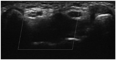
Fig. 2 Bifurcated ulnar nerve in Guyon's canal.
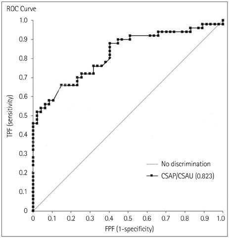
Fig. 3 ROC curve for CSApisiform/CSAulnar. CSAP: median nerve cross-sectional area, CSAU: ulnar nerve cross-sectional area, FPF: false positive fraction, TPF: true positive fraction, ROC: receiver operating characteristic.
Reference
-
1. de Krom MC, Knipschild PG, Kester AD, Thijs CT, Boekkooi PF, Spaans F. Carpal tunnel syndrome: prevalence in the general population. J Clin Epidemiol. 1992; 45:373–376.
Article2. Keith MW, Masear V, Chung KC, Maupin K, Andary M, Amadio PC, et al. American Academy of Orthopaedic Surgeons Clinical Practice Guideline on diagnosis of carpal tunnel syndrome. J Bone Joint Surg Am. 2009; 91:2478–2479.
Article3. Buchberger W, Judmaier W, Birbamer G, Lener M, Schmidauer C. Carpal tunnel syndrome: diagnosis with high-resolution sonography. AJR Am J Roentgenol. 1992; 159:793–798.
Article4. Buchberger W, Schön G, Strasser K, Jungwirth W. High-resolution ultrasonography of the carpal tunnel. J Ultrasound Med. 1991; 10:531–537.
Article5. Practice parameter for carpal tunnel syndrome (summary statement). Report of the Quality Standards Subcommittee of the American Academy of Neurology. Neurology. 1993; 43:2406–2409.6. Beekman R, Visser LH. Sonography in the diagnosis of carpal tunnel syndrome: a critical review of the literature. Muscle Nerve. 2003; 27:26–33.
Article7. Duncan I, Sullivan P, Lomas F. Sonography in the diagnosis of carpal tunnel syndrome. AJR Am J Roentgenol. 1999; 173:681–684.
Article8. Wong SM, Griffith JF, Hui AC, Tang A, Wong KS. Discriminatory sonographic criteria for the diagnosis of carpal tunnel syndrome. Arthritis Rheum. 2002; 46:1914–1921.
Article9. Werner RA, Andary M. Electrodiagnostic evaluation of carpal tunnel syndrome. Muscle Nerve. 2011; 44:597–607.
Article10. Zaidman CM, Al-Lozi M, Pestronk A. Peripheral nerve size in normals and patients with polyneuropathy: an ultrasound study. Muscle Nerve. 2009; 40:960–966.
Article11. Ulaşli AM, Duymuş M, Nacir B, Rana Erdem H, Koşar U. Reasons for using swelling ratio in sonographic diagnosis of carpal tunnel syndrome and a reliable method for its calculation. Muscle Nerve. 2013; 47:396–402.
Article12. Hobson-Webb LD, Massey JM, Juel VC, Sanders DB. The ultrasonographic wrist-to-forearm median nerve area ratio in carpal tunnel syndrome. Clin Neurophysiol. 2008; 119:1353–1357.
Article13. Sarría L, Cabada T, Cozcolluela R, Martínez-Berganza T, García S. Carpal tunnel syndrome: usefulness of sonography. Eur Radiol. 2000; 10:1920–1925.
Article14. Mhoon JT, Juel VC, Hobson-Webb LD. Median nerve ultrasound as a screening tool in carpal tunnel syndrome: correlation of cross-sectional area measures with electrodiagnostic abnormality. Muscle Nerve. 2012; 46:871–878.
Article15. Lee CH, Kim TK, Yoon ES, Dhong ES. Correlation of high-resolution ultrasonographic findings with the clinical symptoms and electrodiagnostic data in carpal tunnel syndrome. Ann Plast Surg. 2005; 54:20–23.
Article16. Keberle M, Jenett M, Kenn W, Reiners K, Peter M, Haerten R, et al. Technical advances in ultrasound and MR imaging of carpal tunnel syndrome. Eur Radiol. 2000; 10:1043–1050.
Article17. Kang S, Kwon HK, Kim KH, Yun HS. Ultrasonography of median nerve and electrophysiologic severity in carpal tunnel syndrome. Ann Rehabil Med. 2012; 36:72–79.
Article18. Lange J. Carpal tunnel syndrome diagnosed using ultrasound as a first-line exam by the surgeon. J Hand Surg Eur Vol. 2013; 38:627–632.
Article19. Bueno-Gracia E, Tricas-Moreno JM, Fanlo-Mazas P, Malo-Urries M, Haddad-Garay M, Estebanez-de-Miguel E, et al. [Relationship between ultrasound measurements of the median nerve and electrophysiological severity in carpal tunnel syndrome]. Rev Neurol. 2015; 61:441–446.20. Visser LH, Smidt MH, Lee ML. Diagnostic value of wrist median nerve cross sectional area versus wrist-to-forearm ratio in carpal tunnel syndrome. Clin Neurophysiol. 2008; 119:2898–2899. author reply 2899.
Article21. Yesildag A, Kutluhan S, Sengul N, Koyuncuoglu HR, Oyar O, Guler K, et al. The role of ultrasonographic measurements of the median nerve in the diagnosis of carpal tunnel syndrome. Clin Radiol. 2004; 59:910–915.
Article22. Eom YI, Choi MH, Kim YK, Joo IS. Sonographic findings in the ulnar nerve according to the electrophysiologic stage of carpal tunnel syndrome. J Ultrasound Med. 2015; 34:1027–1034.
Article23. Ginanneschi F, Filippou G, Reale F, Scarselli C, Galeazzi M, Rossi A. Ultrasonographic and functional changes of the ulnar nerve at Guyon's canal after carpal tunnel release. Clin Neurophysiol. 2010; 121:208–213.
Article
- Full Text Links
-
- Actions
-
Cited
- CITED
-
- Close
- Share
-
- Similar articles
-
- Ultrasonographic Findings of Median Nerve Change according to the Wrist Position
- Reappraisal of Nerve Conduction Studies in Carpal Tunnel Syndrome
- Subclinical Ulnar Neuropathy in Carpal Tunnel Syndrome
- Ultrasonographic Study of Median Nerve after Carpal Tunnel Release
- Ulnar Neuropathy at the Wrist in a Patient with Carpal Tunnel Syndrome after Open Carpal Tunnel Release
