Ulnar Neuropathy at the Wrist in a Patient with Carpal Tunnel Syndrome after Open Carpal Tunnel Release
- Affiliations
-
- 1Department of Physical Medicine and Rehabilitation, College of Medicine, Korea University, Ansan 425-707, Korea. rmkdh@korea.ac.kr
- KMID: 2266772
- DOI: http://doi.org/10.5535/arm.2012.36.2.291
Abstract
- Ulnar neuropathy at the wrist is rarely reported as complications of carpal tunnel release. Since it can sometimes be confused with recurrent median neuropathy at the wrist or ulnar neuropathy at the elbow, an electrodiagnostic study is useful for detecting the lesion in detail. We present a case of a 51-year-old woman with a two-week history of right ulnar palm and 5th digit tingling sensation that began 3 months after open carpal tunnel release surgery of the right hand. Electrodiagnostic tests such as segmental nerve conduction studies of the ulnar nerve at the wrist were useful for localization of the lesion, and ultrasonography helped to confirm the presence of the lesion. After conservative management, patient symptoms were progressively relieved. Combined electrodiagnostic studies and ultrasonography may be helpful for diagnosing and detecting ulnar neuropathies of the wrist following carpal tunnel release surgery.
MeSH Terms
Figure
-
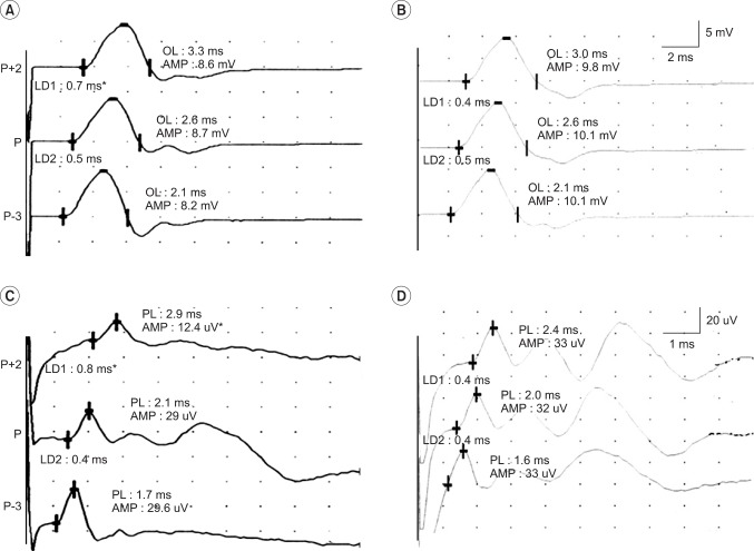
Fig. 1 Initial ulnar motor and sensory short segment studies (SSS) at the wrist (A, C) demonstrate abnormally prolonged differences in latency within the segment between the pisiform (P) and 2 cm proximal to the pisiform (P+2), as well as a conduction block (54.1%) of the ulnar sensory nerve in the same segment. Long-term follow-up SSS (about 4 years later) revealed complete recovery of the focal ulnar nerve lesion (B, D). AMP: Amplitude, P-3: 3 cm distal to the pisiform, LD1: Latency difference (motor, onset latency; sensory, peak latency) in the segment between P and P+2, LD2: Latency difference in the segment between P and P-3. Asterisks indicate abnormal values.
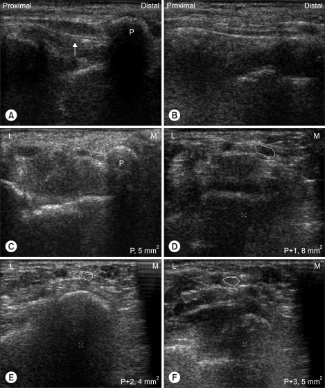
Fig. 2 Longitudinal view of right wrist ultrasonography (A) demonstrates ulnar nerve swelling (arrow) between the pisiform (P) and 2 cm proximal to the P (P+2), as compared to the left side (B). Cross-sectional views (C-F) of the right ulnar nerve reveals a larger area (8 mm2) at the 1 cm proximal to P (P+1, D) than at the other sites (C, E, F; 4-5 mm2). L: Lateral side, M: Medial side, P+3: 3 cm proximal to P.
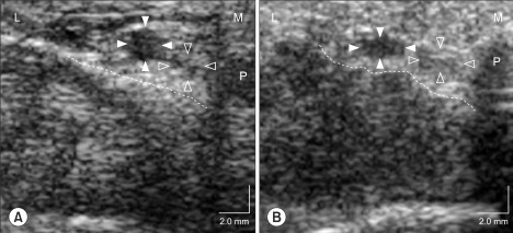
Fig. 3 Ultrasonographic view of Guyon's canal after carpal tunnel release (B) demonstrates the mildly flattened ulnar artery (arrowhead) and nerve (blank arrowhead), and irregular floor line (dotted line) of Guyon's canal compared to the preoperative ultrasonographic view (A).
Reference
-
1. Thurston A, Lam N. Results of open carpal tunnel release: a comprehensive, retrospective study of 188 hands. Aust N Z J Surg. 1997; 67:283–288. PMID: 9152160.
Article2. MacDonald RI, Lichtman DM, Hanlon JJ, Wilson JN. Complications of surgical release for carpal tunnel syndrome. J Hand Surg Am. 1978; 3:70–76. PMID: 621368.
Article3. Pingree MJ, Bosch EP, Liu P, Smith BE. Delayed ulnar neuropathy at the wrist following open carpal tunnel release. Muscle Nerve. 2005; 31:394–397. PMID: 15627268.
Article4. Kim DH, Kang YK, Hwang M, Kwon HK, Lee HJ, Kim BG. Reference values of fractionated neurography of the ulnar nerve at the wrist in healthy subjects. Clin Neurophysiol. 2005; 116:2853–2857. PMID: 16221563.
Article5. Tung TH, Mackinnon SE. Secondary carpal tunnel surgery. Plast Reconstr Surg. 2001; 107:1830–1843. PMID: 11391209.
Article6. Cassvan A, Rosenberg A, Rivera LF. Ulnar nerve involvement in carpal tunnel syndrome. Arch Phys Med Rehabil. 1986; 67:290–292. PMID: 3707312.7. Moghtaderi A, Ghafarpoor M. The dilemma of ulnar nerve entrapment at wrist in carpal tunnel syndrome. Clin Neurol Neurosurg. 2009; 111:151–155. PMID: 19084328.
Article8. Ablove RH, Moy OJ, Peimer CA, Wheeler DR, Diao E. Pressure changes in Guyon's canal after carpal tunnel release. J Hand Surg Br. 1996; 21:664–665. PMID: 9230958.
Article9. Lee CH, Kim TK, Yoon ES, Dhong ES. Postoperative morphologic analysis of carpal tunnel syndrome using high-resolution ultrasonography. Ann Plast Surg. 2005; 54:143–146. PMID: 15655463.
Article10. Wolfe SW, Hotchkiss RN, Pederson WC, Kozin SH. Green's operative hand surgery. 2011. 6th ed. Philadelphia: Elsevier/Churchill Livingston;p. 995.
