Clear Cell Papulosis: A Case Report
- Affiliations
-
- 1Department of Pathology, Asan Medical Center, University of Ulsan College of Medicine, Seoul, Korea. csikpark@amc.seoul.kr
- KMID: 2353605
- DOI: http://doi.org/10.4132/jptm.2016.02.16
Abstract
- No abstract available.
Figure
-
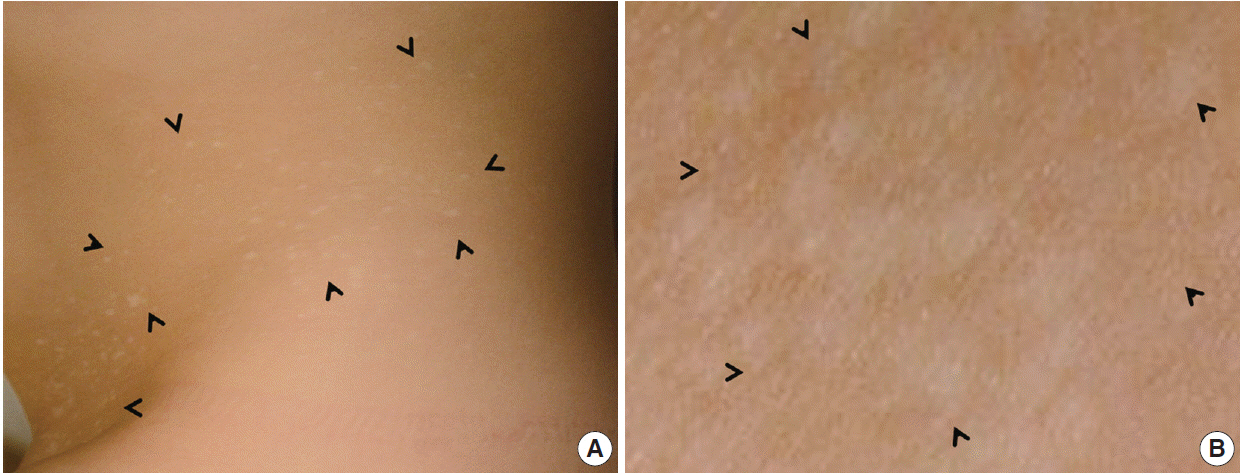
Fig. 1. (A) Many small whitish macules are present on the trunk and are particularly numerous in the lower abdomen and pubic area (arrowheads). The milk-line distribution of the lesions is notable. (B) Close-up view of the right pubic areas showing round-to-oval hypopigmented with no scales (arrowheads).
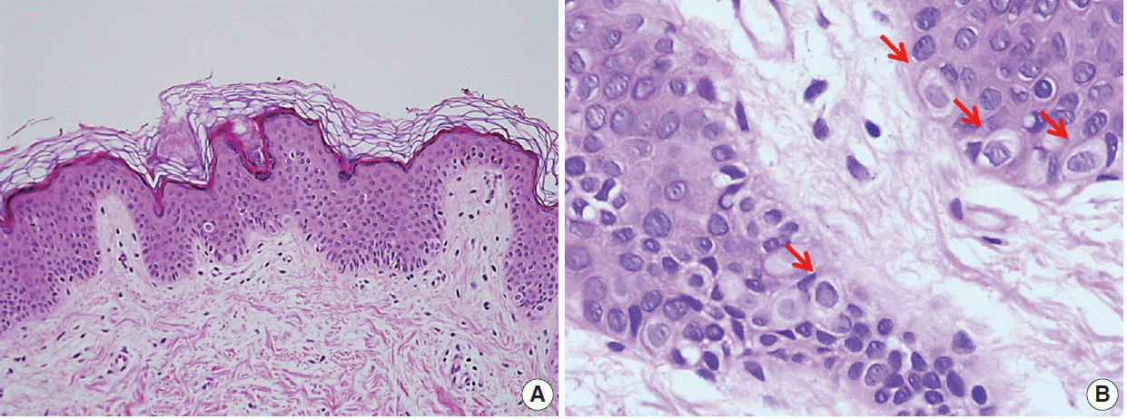
Fig. 2. Biopsy of a clear cell papulosis lesion revealing scattered single clear cells in the basal layer with mild acanthosis and no alteration of the keratin layer. (A) There is minimal inflammatory cell infiltration in the dermis. (B) At higher magnification, solitary benign-appearing pagetoid cells with an abundant clear cytoplasm could be observed in the epidermis. No nuclear atypia is noted (arrows, clear cell).
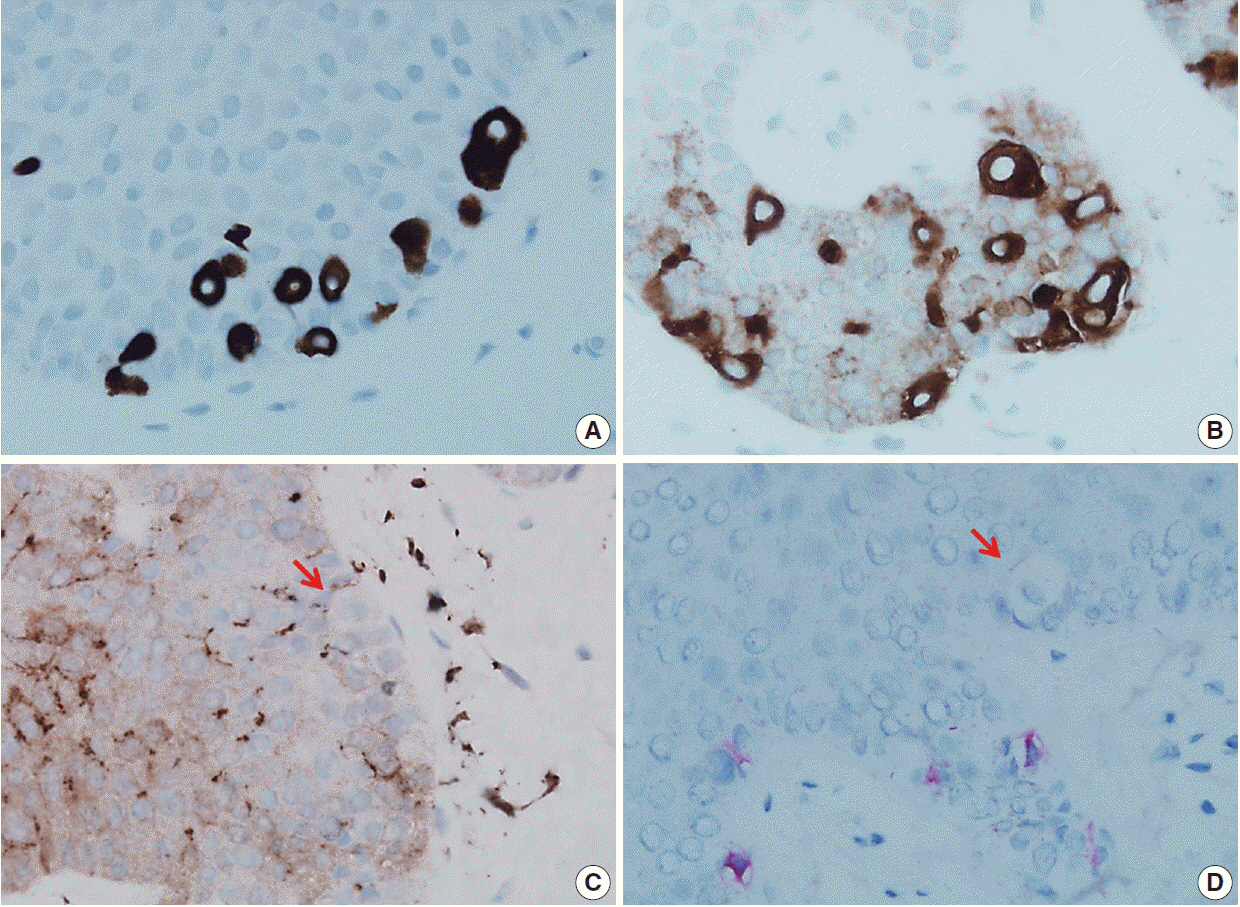
Fig. 3. The pagetoid cells strongly express cytokeratin 7 (A) and carcinoembryonic antigen (B) but are negative for either S100 protein (C) or Melan A (D) (arrows, clear cell).
Reference
-
1. Silverberg NB. Clear cell papulosis. In : Silverberg NB, Duran-McKinster C, Tay YK, editors. Pediatric skin of color. New York: Springer;2015. p. 229–30.2. Kuo TT, Chan HL, Hsueh S. Clear cell papulosis of the skin. A new entity with histogenetic implications for cutaneous Paget’s disease. Am J Surg Pathol. 1987; 11:827–34.3. Mohanty SK, Arora R, Kakkar N, Kumar B. Clear cell papulosis of the skin. Ann Diagn Pathol. 2002; 6:385–8.
Article4. Tseng FW, Kuo TT, Lu PH, Chan HL, Chan MJ, Hui RC. Long-term follow-up study of clear cell papulosis. J Am Acad Dermatol. 2010; 63:266–73.
Article5. Yu Y, Sukhatme S, Loo DS. Clear cell papulosis: a connection of clear cells to toker cells or paget disease. Arch Dermatol. 2009; 145:1066–8.
Article6. Wysong A, Sundram U, Benjamin L. Clear-cell papulosis: a rare entity that may be misconstrued pathologically as normal skin. Pediatr Dermatol. 2012; 29:195–8.
Article7. Chen YH, Wong TW, Lee JY. Depigmented genital extramammary Paget’s disease: a possible histogenetic link to Toker’s clear cells and clear cell papulosis. J Cutan Pathol. 2001; 28:105–8.
Article8. Kim YC, Mehregan DA, Bang D. Clear cell papulosis: an immunohistochemical study to determine histogenesis. J Cutan Pathol. 2002; 29:11–4.
Article9. Di Tommaso L, Franchi G, Destro A, et al. Toker cells of the breast: morphological and immunohistochemical characterization of 40 cases. Hum Pathol. 2008; 39:1295–300.
Article
- Full Text Links
-
- Actions
-
Cited
- CITED
-
- Close
- Share
-
- Similar articles
-
- A Case of Clear Cell Papulosis Misdiagnosed as a Negative for Carcinoembryonic Antigen and Literature Review
- A Case of Lymphomatoid Papulosis Associated with Early Mycosis Fungoides
- A Case of Regional Lymphomatoid Papulosis in Childhood
- Lymphomatoid Papulosis Developed after Remission of Hodgkin's Lymphoma
- Lymphomatoid Papulosis Developing in an Mycosis Fungoides Lesion after Narrow Band UVB Phototherapy and Topical Corticosteroid Application
