Dorsoscapularis triangularis: embryological and phylogenetic characterization of a rare variation of trapezius
- Affiliations
-
- 1Department of Anatomy, Lady Hardinge Medical College and Associated Hospitals, New Delhi, India. dr_lalit_mehra@yahoo.com
- KMID: 2353348
- DOI: http://doi.org/10.5115/acb.2016.49.3.213
Abstract
- The muscle trapezius shows considerable morphological diversity. Variations include an anomalous origin and complete or partial absence of the muscle. The present study reported, a hitherto undocumented complete bilateral absence of the cervical part of trapezius. Based on its peculiar origin and insertion, it was named dorsoscapularis triangularis. The embryological, phylogenetic and molecular basis of the anomaly was elucidated. Failure of cranial migration of the trapezius component of the branchial musculature anlage to gain attachment on the occipital bone, cervical spinous processes, ligamentum nuchae between 11 mm and 16 mm stage of the embryo, resulted in this anomaly. A surgeon operating on the head and neck region or a radiologist analyzing a magnetic resonance imaging of the cervical region would find the knowledge of this morphological variation of trapezius useful in making clinical decisions.
Keyword
MeSH Terms
Figure
-
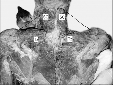
Fig. 1 Dissection of the back and neck showing absence of cervical part of trapezius. Dotted black line depicting the normal extent of the cervical part of trapezius. SC, splenius capitis; Tz, trapezius.
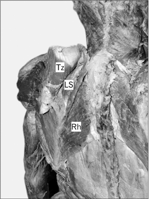
Fig. 2 Muscles undercover of trapezius seen after reflecting it laterally. LS, levator scapulae; Rh, rhomboides; Tz, trapezius.
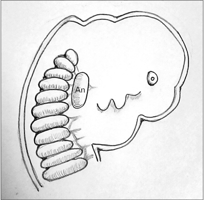
Fig. 3 Diagrammatic representation of a 7 mm embryo showing the Anlage for sternocleidomastoid and trapezius lying ventral to the cranial myotomes. Modified from Keibel and Mall (1910) [8]. An, branchial muscle anlage for sternocleidomastoid and trapezius.
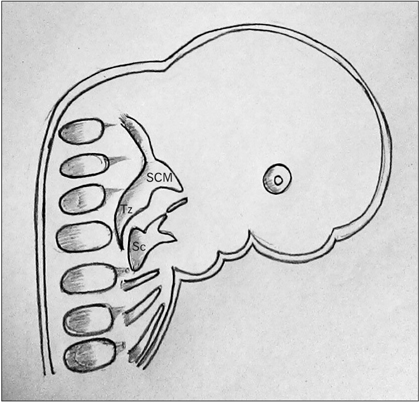
Fig. 4 Diagrammatic representation of an 11 mm embryo showing the splitting of the anlage into ventral sternocleidomastoid and dorsal trapezius. Modified from Keibel and Mall (1910) [8]. Sc, scapula; SCM, sternocleidomastoid; Tz, trapezius.

Fig. 5 Diagrammatic representation of a 16 mm embryo showing failure of cranial migration of the trapezial component to gain attachment on the cervical spinous processes, ligamentum nuchae and the occipital bone. Modified from Keibel and Mall (1910) [8]. De, deltoid; SC, splenius capitis; SCM, sternocleidomastoid; Tz, trapezius.
Cited by 1 articles
-
Bilateral sternocleidomastoid variant with six distinct insertions along the superior nuchal line
Graham Dupont, Joe Iwanaga, Juan J. Altafulla, Stefan Lachkar, Rod J. Oskouian, R. Shane Tubbs
Anat Cell Biol. 2018;51(4):305-308. doi: 10.5115/acb.2018.51.4.305.
Reference
-
1. Standring S. Gray's anatomy: the anatomical basis of clinical practice. 40th ed. Madrid: Elsevier Churchill Livingstone;2008.2. Colletti G, Tewfik K, Bardazzi A, Allevi F, Chiapasco M, Mandalà M, Rabbiosi D. Regional flaps in head and neck reconstruction: a reappraisal. J Oral Maxillofac Surg. 2015; 73:571.e1–571.e10.3. Allouh M, Mohamed A, Mhanni A. Complete unilateral absence of trapezius muscle. McGill J Med. 2004; 8:31–33.4. Horan FT, Bonafede RP. Bilateral absence of the trapezius and sternal head of the pectoralis major muscles: a case report. J Bone Joint Surg Am. 1977; 59:133.5. Yiyit N, Işıtmangil T, Öksüz S. Clinical analysis of 113 patients with Poland syndrome. Ann Thorac Surg. 2015; 99:999–1004.6. Miyamoto RT, Yune HY, Rosevear WH. Klippel-Feil syndrome and associated ear deformities. Am J Otol. 1983; 5:113–119.7. Takahashi H, Umeda M, Sakakibara A, Shigeta T, Minamikawa T, Shibuya Y, Komori T. Absence of the sternocleidomastoid muscle in a patient that underwent neck dissection for squamous cell carcinoma of the tongue. Kobe J Med Sci. 2014; 59:E167–E171.8. Keibel F, Mall FP. Manual of human embryology. Philadelphia, PA: J.B Lippincott Company;1910.9. Diogo R, Wood BA. Comparative anatomy and phylogeny of primate muscles and human evolution. New York: CRC Press;2012.10. Mehta V, Arora J, Kumar A, Nayar AK, Ioh HK, Gupta V, Suri RK, Rath G. Bipartite clavicular attachment of the sternocleidomastoid muscle: a case report. Anat Cell Biol. 2012; 45:66–69.11. Chen WL, Li J, Yang Z, Huang Z, Wang J, Zhang B. Extended vertical lower trapezius island myocutaneous flap in reconstruction of oral and maxillofacial defects after salvage surgery for recurrent oral carcinoma. J Oral Maxillofac Surg. 2007; 65:205–211.12. Umeda M, Shigeta T, Takahashi H, Oguni A, Kataoka T, Minamikawa T, Shibuya Y, Komori T. Shoulder mobility after spinal accessory nerve-sparing modified radical neck dissection in oral cancer patients. Oral Surg Oral Med Oral Pathol Oral Radiol Endod. 2010; 109:820–824.
- Full Text Links
-
- Actions
-
Cited
- CITED
-
- Close
- Share
-
- Similar articles
-
- An Anatomic Variation of the Trapezius Muscle in a Korean: The Cleido-occipitalis Cervicalis
- Keratitis by Acanthamoeba triangularis: Report of Cases and Characterization of Isolates
- The First Case of Cutaneous Acanthamoebiasis Caused by Acanthamoeba triangularis in Korea
- Untrapped: bilateral hypoplasia of the trapezius muscle
- Concomitant Variations in Flexor Digitorum Superficialis: A Case Report
