J Cardiovasc Ultrasound.
2016 Sep;24(3):223-228. 10.4250/jcu.2016.24.3.223.
Myocardial Rotation and Torsion in Child Growth
- Affiliations
-
- 1Division of Pediatric Cardiology, Department of Pediatrics, Yonsei University College of Medicine, Seoul, Korea. lucyeun@yuhs.ac
- KMID: 2353100
- DOI: http://doi.org/10.4250/jcu.2016.24.3.223
Abstract
- BACKGROUND
The speckle tracking echocardiography can benefit to assess the regional myocardial deformations. Although, previous reports suggested no significant change in left ventricular (LV) torsion with aging, there are certain differences in LV rotation at the base and apex. The purpose of this study was to evaluate the change and relationship of LV rotation for torsion with aging in children.
METHODS
Forty healthy children were recruited and divided into two groups of twenty based on whether the children were preschool-age (2-6 years of age) or school-age (7-12 years of age). After obtaining conventional echocardiographic data, apical and basal short axis rotation were assessed with speckle tracking echocardiography. LV rotation in the basal and apical short axis planes was determined using six myocardial segments along the central axis.
RESULTS
Apical and basal LV rotation did not show the statistical difference with increased age between preschool- and school-age children. Apical radial strain showed significant higher values in preschool-age children, especially at the anterior (52.8 ± 17.4% vs. 34.7 ± 23.2%, p < 0.02), lateral (55.8 ± 20.4% vs. 36.1 ± 22.7%, p < 0.02), and posterior segments (57.1 ± 17.6% vs. 38.5 ± 21.7%, p < 0.01). The torsion values did not demonstrate the statistical difference between two groups.
CONCLUSION
This study revealed the tendency of higher rotation values in preschool-age children than in school-age children. The lesser values of rotation and torsion with increased age during childhood warrant further investigation.
MeSH Terms
Figure
-
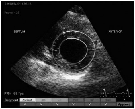
Fig. 1 Measurement process of left ventricular rotation by two-dimensional speckle tracking echocardiographic imaging.

Fig. 2 Rotation data were acquired with speckle tracking echocardiography for (A) basal clockwise rotation, and (B) apical counterclockwise rotation, offline analysis at two-dimensional short axis view.
Reference
-
1. Buckberg GD, Weisfeldt ML, Ballester M, Beyar R, Burkhoff D, Coghlan HC, Doyle M, Epstein ND, Gharib M, Ideker RE, Ingels NB, LeWinter MM, McCulloch AD, Pohost GM, Reinlib LJ, Sahn DJ, Sopko G, Spinale FG, Spotnitz HM, Torrent-Guasp F, Shapiro EP. Left ventricular form and function: scientific priorities and strategic planning for development of new views of disease. Circulation. 2004; 110:e333–e336.2. Notomi Y, Lysyansky P, Setser RM, Shiota T, Popović ZB, Martin-Miklovic MG, Weaver JA, Oryszak SJ, Greenberg NL, White RD, Thomas JD. Measurement of ventricular torsion by two-dimensional ultrasound speckle tracking imaging. J Am Coll Cardiol. 2005; 45:2034–2041.3. McDonald IG. The shape and movements of the human left ventricle during systole. A study by cineangiography and by cineradiography of epicardial markers. Am J Cardiol. 1970; 26:221–230.4. Rademakers FE, Buchalter MB, Rogers WJ, Zerhouni EA, Weisfeldt ML, Weiss JL, Shapiro EP. Dissociation between left ventricular untwisting and filling. Accentuation by catecholamines. Circulation. 1992; 85:1572–1581.5. Gibbons Kroeker CA, Ter Keurs HE, Knudtson ML, Tyberg JV, Beyar R. An optical device to measure the dynamics of apex rotation of the left ventricle. Am J Physiol. 1993; 265(4 Pt 2):H1444–H1449.6. Moon MR, Ingels NB Jr, Daughters GT 2nd, Stinson EB, Hansen DE, Miller DC. Alterations in left ventricular twist mechanics with inotropic stimulation and volume loading in human subjects. Circulation. 1994; 89:142–150.7. Hansen DE, Daughters GT 2nd, Alderman EL, Ingels NB, Stinson EB, Miller DC. Effect of volume loading, pressure loading, and inotropic stimulation on left ventricular torsion in humans. Circulation. 1991; 83:1315–1326.8. Yun KL, Niczyporuk MA, Daughters GT 2nd, Ingels NB Jr, Stinson EB, Alderman EL, Hansen DE, Miller DC. Alterations in left ventricular diastolic twist mechanics during acute human cardiac allograft rejection. Circulation. 1991; 83:962–973.9. Maier SE, Fischer SE, McKinnon GC, Hess OM, Krayenbuehl HP, Boesiger P. Evaluation of left ventricular segmental wall motion in hypertrophic cardiomyopathy with myocardial tagging. Circulation. 1992; 86:1919–1928.10. Buchalter MB, Rademakers FE, Weiss JL, Rogers WJ, Weisfeldt ML, Shapiro EP. Rotational deformation of the canine left ventricle measured by magnetic resonance tagging: effects of catecholamines, ischaemia, and pacing. Cardiovasc Res. 1994; 28:629–635.11. DeAnda A Jr, Komeda M, Nikolic SD, Daughters GT 2nd, Ingels NB, Miller DC. Left ventricular function, twist, and recoil after mitral valve replacement. Circulation. 1995; 92:9 Suppl. II458–II466.12. Gibbons Kroeker CA, Tyberg JV, Beyar R. Effects of load manipulations, heart rate, and contractility on left ventricular apical rotation. An experimental study in anesthetized dogs. Circulation. 1995; 92:130–141.13. Kroeker CA, Tyberg JV, Beyar R. Effects of ischemia on left ventricular apex rotation. An experimental study in anesthetized dogs. Circulation. 1995; 92:3539–3548.14. Knudtson ML, Galbraith PD, Hildebrand KL, Tyberg JV, Beyar R. Dynamics of left ventricular apex rotation during angioplasty: a sensitive index of ischemic dysfunction. Circulation. 1997; 96:801–808.15. Stuber M, Scheidegger MB, Fischer SE, Nagel E, Steinemann F, Hess OM, Boesiger P. Alterations in the local myocardial motion pattern in patients suffering from pressure overload due to aortic stenosis. Circulation. 1999; 100:361–368.16. Nagel E, Stuber M, Lakatos M, Scheidegger MB, Boesiger P, Hess OM. Cardiac rotation and relaxation after anterolateral myocardial infarction. Coron Artery Dis. 2000; 11:261–267.17. Sandstede JJ, Johnson T, Harre K, Beer M, Hofmann S, Pabst T, Kenn W, Voelker W, Neubauer S, Hahn D. Cardiac systolic rotation and contraction before and after valve replacement for aortic stenosis: a myocardial tagging study using MR imaging. AJR Am J Roentgenol. 2002; 178:953–958.18. Fuchs E, Müller MF, Oswald H, Thöny H, Mohacsi P, Hess OM. Cardiac rotation and relaxation in patients with chronic heart failure. Eur J Heart Fail. 2004; 6:715–722.19. Tibayan FA, Rodriguez F, Langer F, Zasio MK, Bailey L, Liang D, Daughters GT, Ingels NB Jr, Miller DC. Alterations in left ventricular torsion and diastolic recoil after myocardial infarction with and without chronic ischemic mitral regurgitation. Circulation. 2004; 110:11 Suppl 1. II109–II114.20. Wagner RF, Smith SW, Sandrik JM, Lopez H. Statistics of speckle in ultrasound B-scans. IEEE Trans Sonics Ultrasonics. 1983; 30:156–163.21. Bohs LN, Trahey GE. A novel method for angle independent ultrasonic imaging of blood flow and tissue motion. IEEE Trans Biomed Eng. 1991; 38:280–286.22. Meunier J, Bertrand M. Ultrasonic texture motion analysis: theory and simulation. IEEE Trans Med Imaging. 1995; 14:293–300.23. Sühling M, Jansen C, Arigovindan M, Buser P, Marsch S, Unser M, Hunziker P. Multiscale motion mapping: a novel computer vision technique for quantitative, objective echocardiographic motion measurement independent of Doppler: first clinical description and validation. Circulation. 2004; 110:3093–3099.24. Notomi Y, Srinath G, Shiota T, Martin-Miklovic MG, Beachler L, Howell K, Oryszak SJ, Deserranno DG, Freed AD, Greenberg NL, Younoszai A, Thomas JD. Maturational and adaptive modulation of left ventricular torsional biomechanics: Doppler tissue imaging observation from infancy to adulthood. Circulation. 2006; 113:2534–2541.25. Stopfkuchen H. Changes of the cardiovascular system during the perinatal period. Eur J Pediatr. 1987; 146:545–549.26. Wulfsohn D, Nyengaard JR, Tang Y. Postnatal growth of cardiomyocytes in the left ventricle of the rat. Anat Rec A Discov Mol Cell Evol Biol. 2004; 277:236–247.27. Siedner S, Krüger M, Schroeter M, Metzler D, Roell W, Fleischmann BK, Hescheler J, Pfitzer G, Stehle R. Developmental changes in contractility and sarcomeric proteins from the early embryonic to the adult stage in the mouse heart. J Physiol. 2003; 548(Pt 2):493–505.28. Lahmers S, Wu Y, Call DR, Labeit S, Granzier H. Developmental control of titin isoform expression and passive stiffness in fetal and neonatal myocardium. Circ Res. 2004; 94:505–513.29. Senzaki H, Akagi M, Hishi T, Ishizawa A, Yanagisawa M, Masutani S, Kobayashi T, Awa S. Age-associated changes in arterial elastic properties in children. Eur J Pediatr. 2002; 161:547–551.30. Oxenham H, Sharpe N. Cardiovascular aging and heart failure. Eur J Heart Fail. 2003; 5:427–434.31. Setser RM, Kasper JM, Lieber ML, Starling RC, McCarthy PM, White RD. Persistent abnormal left ventricular systolic torsion in dilated cardiomyopathy after partial left ventriculectomy. J Thorac Cardiovasc Surg. 2003; 126:48–55.32. Notomi Y, Setser RM, Shiota T, Martin-Miklovic MG, Weaver JA, Popović ZB, Yamada H, Greenberg NL, White RD, Thomas JD. Assessment of left ventricular torsional deformation by Doppler tissue imaging: validation study with tagged magnetic resonance imaging. Circulation. 2005; 111:1141–1147.33. Colan SD, Parness IA, Spevak PJ, Sanders SP. Developmental modulation of myocardial mechanics: age- and growth-related alterations in afterload and contractility. J Am Coll Cardiol. 1992; 19:619–629.34. Harada K, Suzuki T, Shimada K, Takada G. Role of left ventricular mass/volume ratio on transmitral flow velocity patterns from infancy to childhood. Int J Cardiol. 1998; 63:9–14.35. Nidorf SM, Picard MH, Triulzi MO, Thomas JD, Newell J, King ME, Weyman AE. New perspectives in the assessment of cardiac chamber dimensions during development and adulthood. J Am Coll Cardiol. 1992; 19:983–988.
- Full Text Links
-
- Actions
-
Cited
- CITED
-
- Close
- Share
-
- Similar articles
-
- Left Ventricular Rotation and Twist: Why Should We Learn?
- Decreased Left Ventricular Torsion and Untwisting in Children with Dilated Cardiomyopathy
- The Prediction for Neutral Rotation of Tibia by the Image of Contralateral Tibia-Fibula
- A Case of the Torsion of the Term Pregnant Uterus with a Transverse Lie of the Fetus
- Torsion of the Cryptorchid Testis: Report of One Case
