Folliculosebaceous Cystic Hamartoma of the Eyelid
- Affiliations
-
- 1Department of Ophthalmology, Maryknoll Medical Center, Busan, Korea. eyerheu@hanafos.com
- KMID: 2351878
- DOI: http://doi.org/10.3341/jkos.2016.57.9.1460
Abstract
- PURPOSE
Folliculosebaceous cystic hamartoma is a rare cutaneous hamartoma consisting of dilated folliculosebaceous units invested in mesenchymal elements. There is no report of folliculosebaceous cystic hamartoma case occurred in the eyelid. We report here on this case along with a review of the relevant literature.
CASE SUMMARY
72-year-old female visited for the complaint of a mass in right upper eyelid. The mass was 1.9 × 1.2 cm sized and palpated in the subcutaneous level of right upper eyelid. The mass was not tender and had hardness like rubber. It was covered by skin without adhesion but fixed on the upper tarsal plate. Turning the eyelid inside out, it was found that the upper tarsal plate was penetrated by the mass. There was no specific finding except both cataract by other ophthalmic examination. The paranasal sinus computed tomography finding was well demarcated 0.9 cm sized mass with calcification. The excisional biopsy was performed for diagnosis and treatment. In pathologic finding, various sized normal sebaceous lobules were connected with the dilated follicles through the sebaceous canal and formed infundibular structure. There were sclerosing collagen, adipose cells and vessels between follicles and sebaceous lobules. So it was compatible with folliculosebaceous cystic hamartoma. 18 months later, there was no recurrence and wound was clear.
CONCLUSIONS
Folliculosebaceous cystic hamartoma of the eye lid is rare disease, and differential diagnosis is necessary in patient with mass of eyelid.
MeSH Terms
Figure
-

Figure 1. Clinical findings. (A) Clinical picture at presentation. It showed a skin colored mass on the right upper eyelid. (B) Photograph when the eyelid was turned inside out. It showed the upper tarsal plate was penetrated by the mass. (C) Computed tomography. A 0.9 cm sized mass with soft tissue density was seen at right upper eyelid (arrow). (D) Gross picture of the mass. It showed ivory white color with well defined surface and the cutting was ivory white and solid.
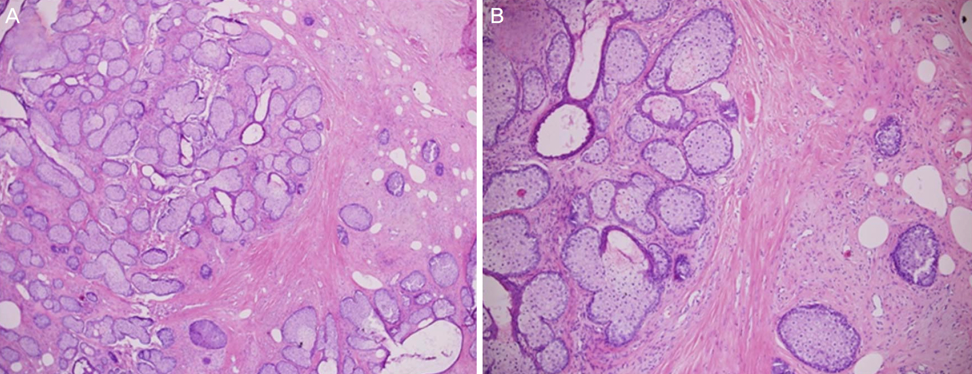
Figure 2. Histopathologic findings. (A) It revealed a numerous mature sebaceous lobules radiating from various sized cystic structures resembling the follicular infundibulum (Hematoxylineosin, ×40). (B) The stroma surrounding the epithelial component of the cyst and sebaceous lobules consisted of densely laminated collagen bundles with adipocytes and small venules (Hematoxylin-eosin, ×100).
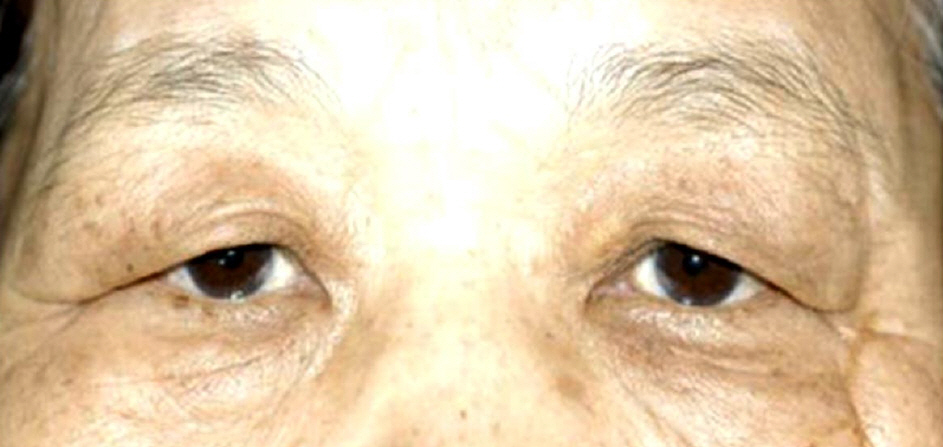
Figure 3. Photograph of the patient 18 months later after treatment. She has remained without recurrence.
Reference
-
References
1. Kimura T, Miyazawa H, Aoyagi T, Ackerman AB. Folliculosebaceous cystic hamartoma: A distinctive malformation of the skin. Am J Dermatopathol. 1991; 13:213–20.2. Ansai S, Kimura T, Kawana S. A clinicoplathologic study of folliculosebaceous cystic hamartoma. Am J Dermatopathol. 2010; 32:815–20.3. Suarez-Pañaranda JM, Vieites B, Ramírez-Santos A, et al. Clinicopathological and immnuohistochemical findings in a series of folliculosebaceous cystic hamartoma. J Cutan Pathol. 2009; 36:251–6.4. El-Darouty MA, Marzouk SA, Abdel-Halim MR, et al. Folliculosebaceous cystic hamartoma. Int J Dermatol. 2001; 40:454–7.
Article5. Ratnakar KS. Pathology of the eye and orbit. London: Jaypee broh-ers medical publishers;1998. p. 186–8.6. Barnes L. Surgical Pathology of the Head and Neck. 3rd ed.Philadelphia: Informa healthcare;2009. p. 1651–3.7. Sturtz DE, Smith DJ, Calderon MS, Fullen DR. Giant folliculosebaceous cystic hamartoma of the upper extremity. J Cutan Pathol. 2004; 31:289–90.
Article8. Hamada M, Kiryu H, Satoh E, et al. A case of genital folliculosebaceous cystic hamartoma with an unique aggregated manifestation. J Dermatol. 2006; 33:191–5.
Article9. Templeton SF. Folliculosebaceous cystic hamartoma: a clinical pathologic study. J Am Acad Dermatol. 1996; 34:77–81.
Article10. Aloi F, Tomasini C, Pippione M. Folliculosebaceous cystic hamartoma with perifollicular mucinosis. Am J Dermatopathol. 1996; 18:58–62.
Article11. Arnab B. Eyelid Tumors: Clinical Evaluation and Reconstruction Techniques. 1st ed.New Delhi: Springer India;2014. 46:p. 118.12. KDA textbook Editing board. Dermatology. 5th ed.Seoul: RyoMoonGak;2008. p. 697–9.
