Clin Endosc.
2016 Jul;49(4):395-398. 10.5946/ce.2015.109.
Successful Removal of a Large Common Bile Duct Stone by Using Direct Peroral Cholangioscopy and Laser Lithotripsy in a Patient with Severe Kyphosis
- Affiliations
-
- 1Department of Internal Medicine, Wonkwang University School of Medicine, Iksan, Korea. kth@wonkwang.ac.kr
- KMID: 2348262
- DOI: http://doi.org/10.5946/ce.2015.109
Abstract
- A 75-year-old woman with hypertension presented with acute suppurative cholangitis. Chest radiography revealed severe kyphosis. Abdominal computed tomography revealed a large stone impacted in the common bile duct (CBD). The patient underwent emergent endoscopic retrograde cholangiopancreatography, and cholangiography revealed a large stone (7×3 cm) in the CBD that could not be captured using a large basket. We could not use the percutaneous approach for stone fragmentation by using a cholangioscope because of severe degenerative kyphosis. Finally, we performed holmium laser lithotripsy under peroral cholangioscopy by using an ultraslim endoscope, and the large stone in the CBD was successfully fragmented and removed without complications.
Keyword
MeSH Terms
Figure
-

Fig. 1. A chest radiograph showing severe kyphosis.
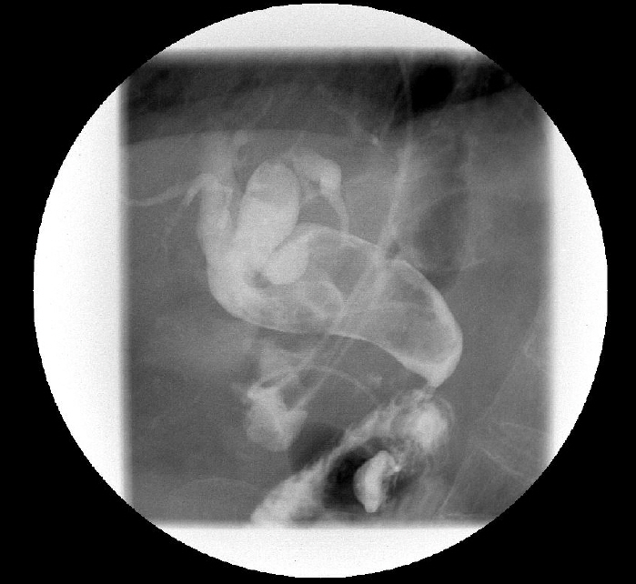
Fig. 2. A cholangiogram showing a large filling defect that suggests a large stone in the common bile duct.
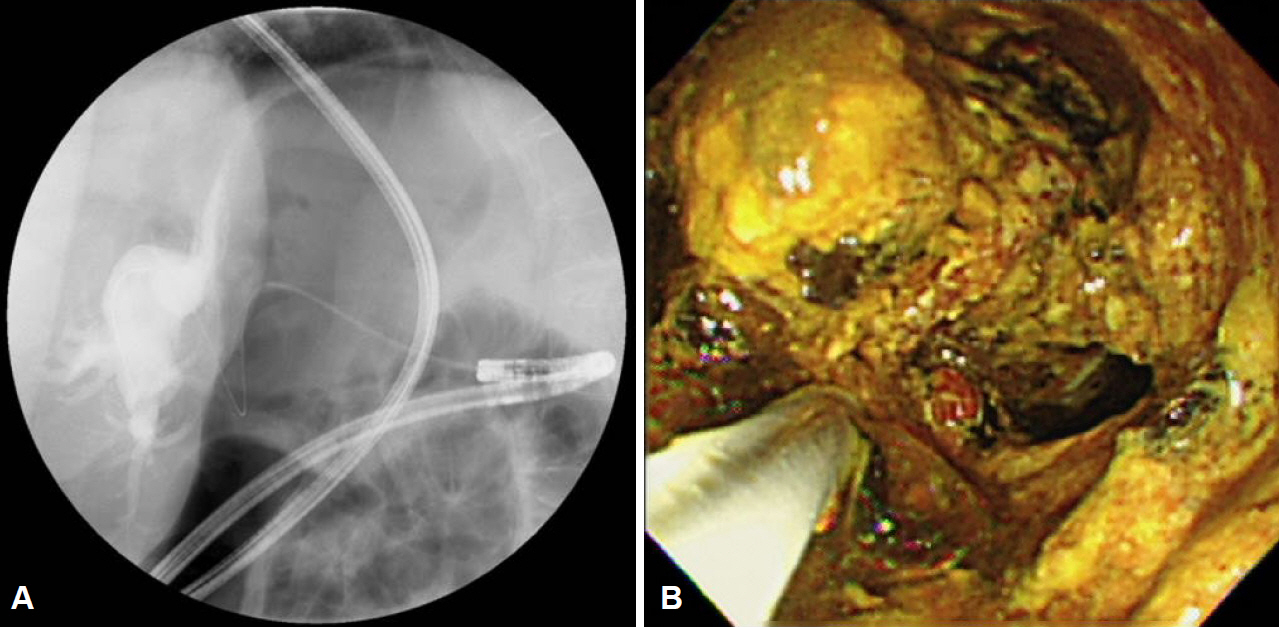
Fig. 3. A radiograph demonstrating peroral direct cholangioscopy. (A) Radiographic view showing the ultraslim endoscope in the common bile duct. (B) Endoscopic view showing stone fragmentation using holmium laser lithotripsy.
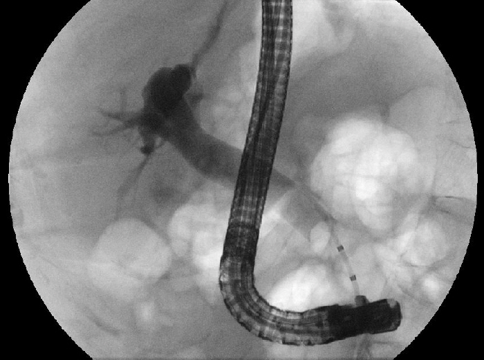
Fig. 4. A balloon cholangiogram showing no stones in the common bile duct.
Reference
-
1. Binmoeller KF, Brückner M, Thonke F, Soehendra N. Treatment of difficult bile duct stones using mechanical, electrohydraulic and extracorporeal shock wave lithotripsy. Endoscopy. 1993; 25:201–206.
Article2. Katanuma A, Maguchi H, Osanai M, Takahashi K. Endoscopic treatment of difficult common bile duct stones. Dig Endosc. 2010; 22 Suppl 1:S90–S97.
Article3. Njeze GE. Gallstones. Niger J Surg. 2013; 19:49–55.4. Trikudanathan G, Navaneethan U, Parsi MA. Endoscopic management of difficult common bile duct stones. World J Gastroenterol. 2013; 19:165–173.
Article5. Moon JH, Ko BM, Choi HJ, et al. Direct peroral cholangioscopy using an ultra-slim upper endoscope for the treatment of retained bile duct stones. Am J Gastroenterol. 2009; 104:2729–2733.
Article6. Lee HJ, Kim SB, Shin CM, et al. A comparison of endoscopic treatments in rectal carcinoid tumors. Surg Endosc. 2016; 30:3491–3498.
Article7. Sola-Vera J, Uceda F, Cuesta R, Vázquez N. Direct peroral cholangioscopy using an ultrathin endoscope: making technique easier. Rev Esp Enferm Dig. 2014; 106:30–36.
Article8. Kim SK, Kang SY, Youn HJ, Jung SH. Comparison of conventional thyroidectomy and endoscopic thyroidectomy via axillo-bilateral breast approach in papillary thyroid carcinoma patients. Surg Endosc. 2016; 30:3419–3425.
Article9. Jiang H, Wu Z, Ding Q, Zhang Y. Ureteroscopic treatment of ureteral calculi with holmium: YAG laser lithotripsy. J Endourol. 2007; 21:151–154.
Article10. Sofer M, Watterson JD, Wollin TA, Nott L, Razvi H, Denstedt JD. Holmium:YAG laser lithotripsy for upper urinary tract calculi in 598 patients. J Urol. 2002; 167:31–34.11. Kim TH, Oh HJ, Choi CS, Yeom DH, Choi SC. Clinical usefulness of transpapillary removal of common bile duct stones by frequency doubled double pulse Nd:YAG laser. World J Gastroenterol. 2008; 14:2863–2866.
Article12. Kim HJ, Ahn JC, Hong SN, Lee WH, Kim JW. Posterior fontanelle approach for uncinectomy and middle meatal antrostomy in endoscopic sinus surgery. Laryngoscope. 2016; 126:1311–1314.
Article13. Chen YK, Pleskow DK. SpyGlass single-operator peroral cholangiopancreatoscopy system for the diagnosis and therapy of bile-duct disorders: a clinical feasibility study (with video). Gastrointest Endosc. 2007; 65:832–841.
Article
- Full Text Links
-
- Actions
-
Cited
- CITED
-
- Close
- Share
-
- Similar articles
-
- Removal of a Large, Intractable Common Bile Duct Stone by Direct Peroral Cholangioscopy Using Upper Gastrointestinal Endoscopy and Polypectomy Snare
- Utility of Direct Peroral Cholangioscopy Using a Multibending Ultraslim Endoscope for Difficult Common Bile Duct Stones
- Successful Endoscopic Treatment of Difficult Common Bile Duct Stones Using Various Interventional Techniques: A Case Report
- Cracking Difficult Biliary Stones
- How Should Biliary Stones be Managed?
