Yonsei Med J.
2015 Nov;56(6):1656-1662. 10.3349/ymj.2015.56.6.1656.
Limb Lengthening in Patients with Achondroplasia
- Affiliations
-
- 1Institute for Rare Diseases and Department of Orthopedic Surgery, Korea University Medical Center, Guro Hospital, Seoul, Korea. songhae@korea.ac.kr
- KMID: 2345897
- DOI: http://doi.org/10.3349/ymj.2015.56.6.1656
Abstract
- PURPOSE
Although bilateral lower-limb lengthening has been performed on patients with achondroplasia, the outcomes for the tibia and femur in terms of radiographic parameters, clinical results, and complications have not been compared with each other. We proposed 1) to compare the radiological outcomes of femoral and tibial lengthening and 2) to investigate the differences of complications related to lengthening.
MATERIALS AND METHODS
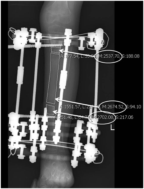
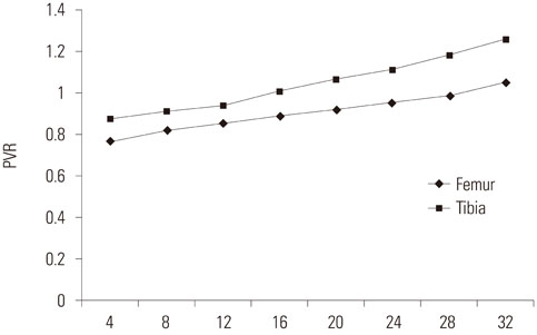
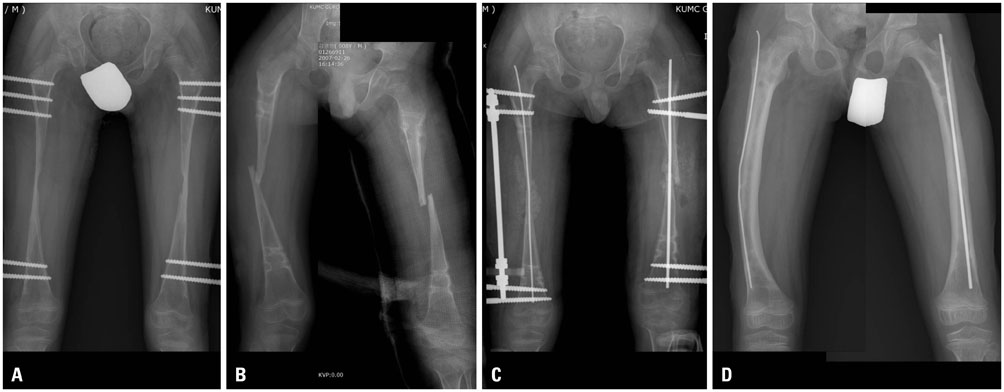
We retrospectively reviewed 28 patients (average age, 14 years 4 months) with achondroplasia who underwent bilateral limb lengthening between 2004 and 2012. All patients first underwent bilateral tibial lengthening, and at 9-48 months (average, 17.8 months) after this procedure, bilateral femoral lengthening was performed. We analyzed the pixel value ratio (PVR) and characteristics of the callus of the lengthened area on serial radiographs. The external fixation index (EFI) and healing index (HI) were computed to compare tibial and femoral lengthening. The complications related to lengthening were assessed.
RESULTS
The average gain in length was 8.4 cm for the femur and 9.8 cm for the tibia. The PVR, EFI, and HI of the tibia were significantly better than those of the femur. Fewer complications were found during the lengthening of the tibia than during the lengthening of the femur.
CONCLUSION
Tibial lengthening had a significantly lower complication rate and a higher callus formation rate than femoral lengthening. Our findings suggest that bilateral limb lengthening (tibia, followed by femur) remains a reasonable option; however, we should be more cautious when performing femoral lengthening in selected patients.
Keyword
MeSH Terms
Figure
Cited by 1 articles
-
Surgical Results of Limb Lengthening at the Femur, Tibia, and Humerus in Patients with Achondroplasia
Kyung Rae Ko, Jong Sup Shim, Chae Hoon Chung, Joo Hwan Kim
Clin Orthop Surg. 2019;11(2):226-232. doi: 10.4055/cios.2019.11.2.226.
Reference
-
1. Takken T, van Bergen MW, Sakkers RJ, Helders PJ, Engelbert RH. Cardiopulmonary exercise capacity, muscle strength, and physical activity in children and adolescents with achondroplasia. J Pediatr. 2007; 150:26–30.2. Paley D, Chaudray M, Pirone AM, Lentz P, Kautz D. Treatment of malunions and mal-nonunions of the femur and tibia by detailed preoperative planning and the Ilizarov techniques. Orthop Clin North Am. 1990; 21:667–691.3. Tandon A, Bhargava SK, Goel S, Bhatt S. Pseudoachondroplasia: a rare cause of rhizomelic dwarfism. Indian J Orthop. 2008; 42:477–479.
Article4. Cattaneo R, Villa A, Catagni M, Tentori L. Limb lengthening in achondroplasia by Ilizarov's method. Int Orthop. 1988; 12:173–179.
Article5. Coleman SS. Simultaneous femoral and tibial lengthening for limb length discrepancies. Arch Orthop Trauma Surg. 1985; 103:359–366.
Article6. Tesiorowski M, Zarzycka M, Kacki W, Jasiewicz B. [Bilateral simultaneous limb lengthening of patients with short stature using the Ilizarov method]. Chir Narzadow Ruchu Ortop Pol. 2002; 67:421–426.7. Paley D. Problems, obstacles, and complications of limb lengthening by the Ilizarov technique. Clin Orthop Relat Res. 1990; 81–104.
Article8. Trivella G, Aldegheri R. Surgical correction of short stature. Acta Paediatr Scand Suppl. 1988; 347:141–146.9. De Bastiani G, Aldegheri R, Trivella G, Renzi-Brivio L, Agostini S, Lavini F. Lengthening of the lower limbs in achondroplastics. Basic Life Sci. 1988; 48:353–355.
Article10. Peretti G, Memeo A, Paronzini A, Marzorati S. Staged lengthening in the prevention of dwarfism in achondroplastic children: a preliminary report. J Pediatr Orthop B. 1995; 4:58–64.
Article11. Li R, Saleh M, Yang L, Coulton L. Radiographic classification of osteogenesis during bone distraction. J Orthop Res. 2006; 24:339–347.12. Zhao L, Fan Q, Venkatesh KP, Park MS, Song HR. Objective guidelines for removing an external fixator after tibial lengthening using pixel value ratio: a pilot study. Clin Orthop Relat Res. 2009; 467:3321–3326.
Article13. Singh S, Song HR, Venkatesh KP, Modi HN, Park MS, Jang KM, et al. Analysis of callus pattern of tibia lengthening in achondroplasia and a novel method of regeneration assessment using pixel values. Skeletal Radiol. 2010; 39:261–266.
Article14. Young JW, Kovelman H, Resnik CS, Paley D. Radiologic assessment of bones after Ilizarov procedures. Radiology. 1990; 177:89–93.
Article15. Babatunde OM, Fragomen AT, Rozbruch SR. Noninvasive quantitative assessment of bone healing after distraction osteogenesis. HSS J. 2010; 6:71–78.
Article16. Hazra S, Song HR, Biswal S, Lee SH, Lee SH, Jang KM, et al. Quantitative assessment of mineralization in distraction osteogenesis. Skeletal Radiol. 2008; 37:843–847.
Article17. Ilizarov GA. The tension-stress effect on the genesis and growth of tissues: Part II. The influence of the rate and frequency of distraction. Clin Orthop Relat Res. 1989; 263–285.18. Kim SJ, Balce GC, Agashe MV, Song SH, Song HR. Is bilateral lower limb lengthening appropriate for achondroplasia?: midterm analysis of the complications and quality of life. Clin Orthop Relat Res. 2012; 470:616–621.
Article19. Shim JS, Chung KH, Ahn JM. Value of measuring bone density serial changes on a picture archiving and communication systems (PACS) monitor in distraction osteogenesis. Orthopedics. 2002; 25:1269–1272.
Article20. Monticelli G, Spinelli R. Leg lengthening by closed metaphyseal corticotomy. Ital J Orthop Traumatol. 1983; 9:139–150.21. Donnan LT, Saleh M, Rigby AS. Acute correction of lower limb deformity and simultaneous lengthening with a monolateral fixator. J Bone Joint Surg Br. 2003; 85:254–260.
Article22. Catagni M. Imaging techniques: the radiographic classification of bone regenerate during distraction. London: Williams and Wilkins;1991.23. Shyam AK, Singh SU, Modi HN, Song HR, Lee SH, An H. Leg lengthening by distraction osteogenesis using the Ilizarov apparatus: a novel concept of tibia callus subsidence and its influencing factors. Int Orthop. 2009; 33:1753–1759.
Article24. Song SH, Sinha S, Kim TY, Park YE, Kim SJ, Song HR. Analysis of corticalization using the pixel value ratio for fixator removal in tibial lengthening. J Orthop Sci. 2011; 16:177–183.
Article25. Yun AG, Severino R, Reinker K. Attempted limb lengthenings beyond twenty percent of the initial bone length: results and complications. J Pediatr Orthop. 2000; 20:151–159.
Article26. Al Turk M HF, Dwiri M. Limb Salvage Procedure in Malignant Bone Tumors using Resection-Shortening-Distraction Technique. A Report of Two Cases. Jordan Royal Med Serve J. 2004; 11:52–55.27. Yasui N, Kawabata H, Kojimoto H, Ohno H, Matsuda S, Araki N, et al. Lengthening of the lower limbs in patients with achondroplasia and hypochondroplasia. Clin Orthop Relat Res. 1997; 298–306.
Article28. Paley D. Current techniques of limb lengthening. J Pediatr Orthop. 1988; 8:73–92.
Article29. Yoshino A, Takao M, Innami K, Matsushita T. Equinus deformity during tibial lengthening with ankle orthoses for equalization of leg-length discrepancies. J Orthop Sci. 2011; 16:756–759.
Article30. Lehman WB, Grant AD, Atar D. Preventing and overcoming equinus contractures during lengthening of the tibia. Orthop Clin North Am. 1991; 22:633–641.
Article31. Cattaneo R, Catagni MA, Guerreschi F. Applications of the Ilizarov method in the humerus. Lengthenings and nonunions. Hand Clin. 1993; 9:729–739.32. Guidera KJ, Hess WF, Highhouse KP, Ogden JA. Extremity lengthening: results and complications with the Orthofix system. J Pediatr Orthop. 1991; 11:90–94.33. Dahl MT, Gulli B, Berg T. Complications of limb lengthening. A learning curve. Clin Orthop Relat Res. 1994; 10–18.
Article
- Full Text Links
- Actions
-
Cited
- CITED
-
- Close
- Share
- Similar articles
-
- Surgical Results of Limb Lengthening at the Femur, Tibia, and Humerus in Patients with Achondroplasia
- Classification and Evaluation of the Callus in Limb Lengthening
- Detection of the Proliferation of Muscle Fibers During Limb Lengthening by Monoclonal Antibody to Bromodeoxyuridine
- A Case of Achondroplasia
- A Case of Achondroplasia


