Comparison of Diffusion Weighted Imaging and Contrast Enhanced Imaging on MRI for Breast Lesions
- Affiliations
-
- 1Department of Radiology, The Catholic University of Korea, Korea. lionmain@catholic.ac.kr
- 2Department of Preventive Medicine, The Catholic University of Korea, Korea.
Abstract
- PURPOSE
To retrospectively compare the MRI findings of diffusion-weighted imaging (DWI) and contrast-enhanced imaging (CEI) of breast lesions.
MATERIALS AND METHODS
From June 2005 to June 2007, 159 patients with 159 biopsy-proven breast lesions underwent breast MRIs including DWI and CEI. Two breast imaging radiologists reviewed the MRI findings by consensus for the presence of a high signal intensity on DWI and enhancement on CEI. The MRI findings and concordance of DWI and CEI findings were correlated with the pathologic diagnosis.
RESULTS
Of the 159 lesions 141 were malignant and 18 were benign. The sensitivity and specificity of DWI for detecting breast cancer was 96.5% (135/141) and 21.4% (5/18), respectively, compared to CEI, whose detection rate was 99.3% (140/141) and 33.3% (6/18), respectively. Larger lesions (> 1 cm) were more frequently detected by DWI. Of the enhanced malignancy, 96.4% (135/140) showed high signal intensity on DWI. The concordance of DWI and CEI findings in malignant lesions was significantly higher than benign lesions (95.7% vs. 61.1%, p<0.0001).
CONCLUSION
High signal intensity on diffuse-weighted MRI shows a high degree of sensitivity for detecting breast cancer.
MeSH Terms
Figure
-

Fig. 1 Invasive ductal carcinoma of 56-year-old women. A. Contrast enhanced axial T1-weighted image shows an enhancing mass with irregular margin in left breast. B. Sagittal contrast enhanced T1-weighted image of left breast shows a rim-enhancement of the mass. C. Diffusion weighted image shows the mass as high-signal intensity mass in left breast.

Fig. 2 A 0.9 cm invasive ductal carcinoma of 49-year-old women. A. Contrast enhanced axial T1-weighted image shows a small enhancing mass in right breast (arrow). B. Diffusion weighted image shows a high-signal intensity focus in right breast (arrow).
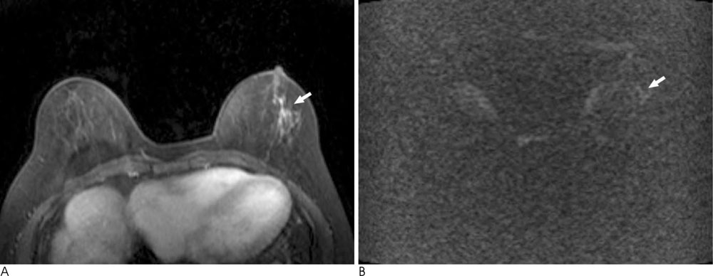
Fig. 3 Ductal carcinoma in situ of 65-year-old women. A. Contrast enhanced axial T1-weighted image show a nonmass enhancement with segmental distribution in left breast (arrow). B. Diffusion weighted image demonstrated no high signal intensity in left breast (arrow).
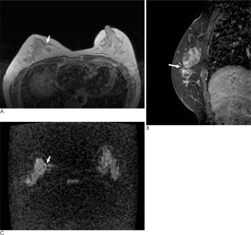
Fig. 4 Ductal carcinoma in situ of 46-year-old women. A, B. Contrast enhanced axial and sagittal T1-weighted images show architectural distortion without enhancement in right breast (arrows). C. The lesion demonstrated focal high signal intensity on diffusion weighted image (arrow).
Reference
-
1. Kriege M, Brekelmans CT, Boetes C, Besnard PE, Zonderland HM, Obdeijn IM, et al. Efficacy of MRI and mammography for breast-cancer screening in women with a familial or genetic predisposition. N Engl J Med. 2004; 351:427–437.2. Teifke A, Behr O, Schmidt M, Victor A, Vomweg TW, Thelen M, et al. Dynamic MR imaging of breast lesions: correlation with microvessel distribution pattern and histologic characteristics of prognosis. Radiology. 2006; 239:351–360.3. Kinkel K, Helbich TH, Esserman LJ, Barclay J, Schwerin EH, Sickles EA, et al. Dynamic high-spatial-resolution MR imaging of suspicious breast lesions: diagnostic criteria and interobserver variability. AJR Am J Roentgenol. 2000; 175:35–43.4. Daniel BL, Yen YF, Glover GH, Ikeda DM, Birdwell RL, Sawyer-Glover AM, et al. Breast disease: dynamic spiral MR imaging. Radiology. 1998; 209:499–509.5. Buadu LD, Murakami J, Murayama S, Hashiguchi N, Sakai S, Masuda K, et al. Breast lesions: correlation of contrast medium enhancement patterns on MR images with histopathologic findings and tumor angiogenesis. Radiology. 1996; 200:639–649.6. Orel SG, Schnall MD, LiVolsi VA, Troupin RH. Suspicious breast lesions: MR imaging with radiologic-pathologic correlation. Radiology. 1994; 190:485–493.7. Kuhl CK, Schrading S, Leutner CC, Morakkabati-Spitz N, Wardelmann E, Fimmers R, et al. Mammography, breast ultrasound, and magnetic resonance imaging for surveillance of women at high familial risk for breast cancer. J Clin Oncol. 2005; 23:8469–8476.8. Yoshikawa MI, Ohsumi S, Sugata S, Kataoka M, Takashima S, Kikuchi K, et al. Comparison of breast cancer detection by diffusion-weighted magnetic resonance imaging and mammography. Radiat Med. 2007; 25:218–223.9. Woodhams R, Matsunaga K, Iwabuchi K, Kan S, Hata H, Kuranami M, et al. Diffusion-weighted imaging of malignant breast tumors: the usefulness of apparent diffusion coefficient (ADC) value and ADC map for the detection of malignant breast tumors and evaluation of cancer extension. J Comput Assist Tomogr. 2005; 29:644–649.10. Lo GG, Ai V, Chan JK, Li KW, Cheung PS, Wong TT, et al. Diffusion-weighted magnetic resonance imaging of breast lesions: first experiences at 3T. J Comput Assist Tomogr. 2009; 33:63–69.11. Park MJ, Cha ES, Kang BJ, Ihn YK, Baik JH. The role of diffusion-weighted imaging and the apparent diffusion coefficient (ADC) values for breast tumors. Korean J Radiol. 2007; 8:390–396.12. Kinoshita T, Yashiro N, Ihara N, Funatu H, Fukuma E, Narita M. Diffusion-weighted half-Fourier single-shot turbo spin echo imaging in breast tumors: differentiation of invasive ductal carcinoma from fibroadenoma. J Comput Assist Tomogr. 2002; 26:1042–1046.13. Charles-Edwards EM, deSouza NM. Diffusion-weighted magnetic resonance imaging and its application to cancer. Cancer Imaging. 2006; 6:135–143.14. Shinya S, Sasaki T, Nakagawa Y, Guiquing Z, Yamamoto F, Yamashita Y. The usefulness of diffusion-weighted imaging (DWI) for the detection of gastric cancer. Hepatogastroenterology. 2007; 54:1378–1381.15. Bakir B, Salmaslioglu A, Poyanli A, Rozanes I, Acunas B. Diffusion weighted MR imaging of pancreatic islet cell tumors. Eur J Radiol. 2009; Forthcoming.16. Abou-El-Ghar ME, El-Assmy A, Refaie HF, El-Diasty T. Bladder cancer: diagnosis with diffusion-weighted MR imaging in patients with gross hematuria. Radiology. 2009; 251:415–442.17. Lin G, Ng KK, Chang CJ, Wang JJ, Ho KC, Yen TC, et al. Myometrial invasion in endometrial cancer: diagnostic accuracy of diffusion-weighted 3.0-T MR imaging--initial experience. Radiology. 2009; 250:784–792.18. Vandecaveye V, De Keyzer F, Vander Poorten V, Dirix P, Verbeken E, Nuyts S, et al. Head and neck squamous cell carcinoma: value of diffusion-weighted MR imaging for nodal staging. Radiology. 2009; 251:134–146.19. Lee KC, Moffat BA, Schott AF, Layman R, Ellingworth S, Juliar R, et al. Prospective early response imaging biomarker for neoadjuvant breast cancer chemotherapy. Clin Cancer Res. 2007; 13(2 Pt 1):443–450.20. Hatakenaka M, Soeda H, Yabuuchi H, Matsuo Y, Kamitani T, Oda Y, et al. Apparent diffusion coefficients of breast tumors: clinical application. Magn Reson Med Sci. 2008; 7:23–29.21. Marini C, Iacconi C, Giannelli M, Cilotti A, Moretti M, Bartolozzi C. Quantitative diffusion-weighted MR imaging in the differential diagnosis of breast lesion. Eur Radiol. 2007; 17:2646–2655.22. Guo Y, Cai YQ, Cai ZL, Gao YG, An NY, Ma L, et al. Differentiation of clinically benign and malignant breast lesions using diffusion-weighted imaging. J Magn Reson Imaging. 2002; 16:172–178.23. Pickles MD, Gibbs P, Lowry M, Turnbull LW. Diffusion changes precede size reduction in neoadjuvant treatment of breast cancer. Magn Reson Imaging. 2006; 24:843–847.24. Kuroki-Suzuki S, Kuroki Y, Nasu K, Nawano S, Moriyama N, Okazaki M. Detecting breast cancer with non-contrast MR imaging: combining diffusion-weighted and STIR imaging. Magn Reson Med Sci. 2007; 6:21–27.
- Full Text Links
-
- Actions
-
Cited
- CITED
-
- Close
- Share
-
- Similar articles
-
- Abbreviated Breast Magnetic Resonance Imaging: Background, Evidence From Studies, and Future Considerations
- The Role of Diffusion-Weighted Imaging and the Apparent Diffusion Coefficient (ADC) Values for Breast Tumors
- Diffusion-Weighted Imaging as a Stand-Alone Breast Imaging Modality
- Diagnosis of Infectious Spondylitis Using Non-Contrast Enhanced MRI With Axial Diffusion-Weighted Images: Comparison With Gadolinium-Enhanced MRI
- Utilization of Magnetic Resonance Imaging in the Diagnosis of Thymic Diseases
