A Case Report of Breast Sparganosis in a Patient with Ipsilateral Breast Cancer: MRI and Ultrasonographic Findings
- Affiliations
-
- 1Department of Radiology, Pusan National University Hospital, Pusan National University School of Medicine and Medical Research Institute, Korea.
- 2Department of Radiology, Pusan National University Yangsan Hospital, Pusan National University School of Medicine and Medical Research Institute, Korea. kschoo0618@naver.com
- 3Department of General Surgery, Pusan National University Hospital, Pusan National University School of Medicine and Medical Research Institute, Korea.
- 4Department of Pathology, Pusan National University Hospital, Pusan National University School of Medicine and Medical Research Institute, Korea.
Abstract
- Sparganosis of the breast is a quite rare parasitic infection of humans and presents as soft tissue masses that mimic breast malignancy or benign tumor, such as fibroadenoma. We present here a case of histologically confirmed breast sparganosis in the upper inner quadrant of the right breast with coexisting breast cancer in the ipsilateral breast upper outer quadrant. Ultrasonography of breast sparganosis showed a well defined, tubular hypoechoic mass with discrete multilayered wall and tubule-in tubule appearance, surrounded by heterogenous hyperechoic areas in the subcutaneous fat layer of the breast. MRI revealed an elongated tubular structure with persistent and progressive enhancement. This is the second report concerned with the MRI and ultrasonographic findings of breast sparganosis and the first report of breast sparganosis in a patient with ipsilateral breast cancer.
MeSH Terms
Figure
-
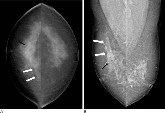
Fig. 1 A 56-year-old woman with two palpable masses in the right breast. Cranio-caudal (A) and medio-lateral oblique (B) mammographic views of the right breast show a focal asymmetry with a well defined cord-like isodense structure (white arrows) without microcalcification in the right breast upper inner quadrant. The black arrow indicates incidental benign punctate calcifications in the right breast upper outer quadrant.
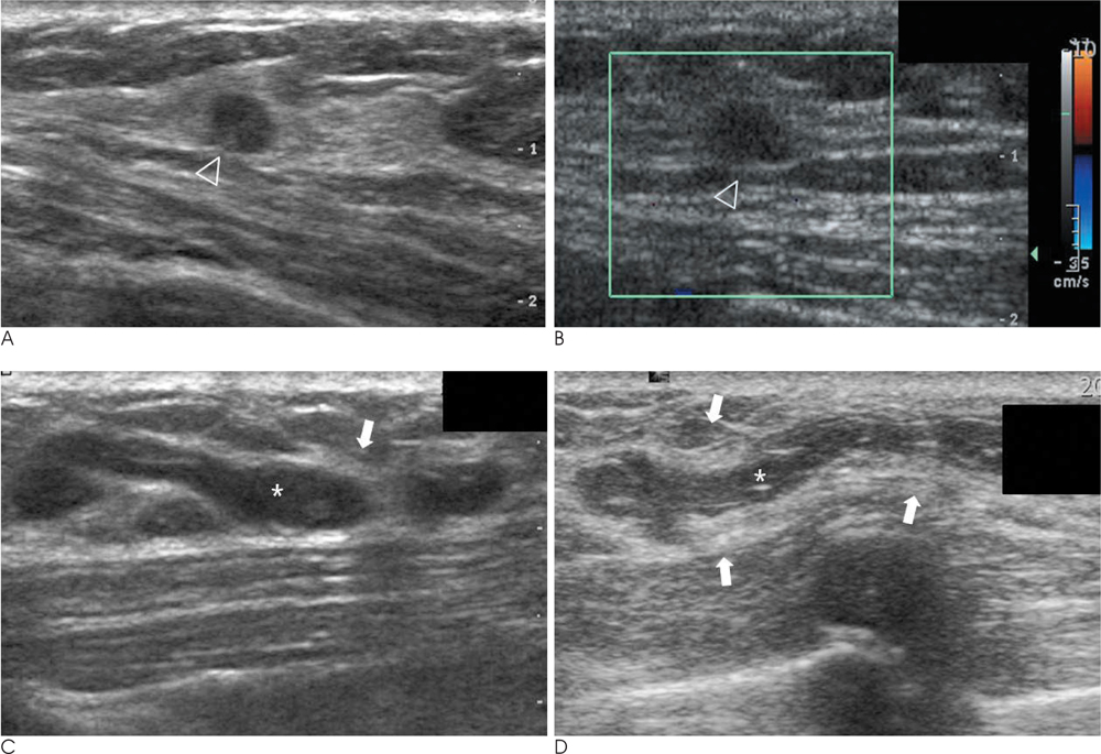
Fig. 2 A 56-year-old woman with coexisting invasive ductal carcinoma (A, B) and breast sparganosis (C, D) in the right breast. A, B. Ultrasound images show a 9 mm sized oval shaped, indistinct margined, hypoechoic mass (arrowhead) in the 10 o'clock position of the right breast upper outer quadrant. This lesion was not apparent on the images of the mammography. Based on the US imaging findings, it is thought to be classified as Breast Imaging Reporting and Data System (BI-RADS) category 4a. C, D. Ultrasound images show a well defined tubular hypoechoic mass with discrete multilayered wall and tubule-in tubule appearance (asterisk), surrounded by heterogenous hyperechoic areas of the subcutaneous fat layer (arrows) in the 2 o'clock position of the ipsilateral right breast upper inner quadrant.
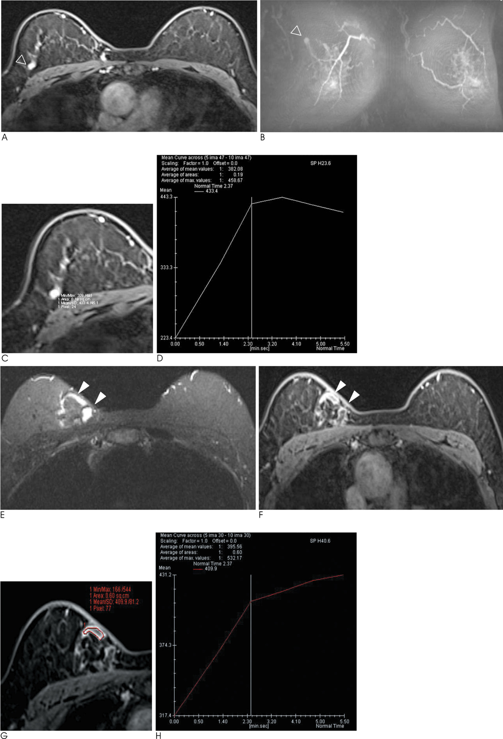
Fig. 3 A 56-year-old woman with coexisting invasive ductal carcinoma (A-D) and breast sparganosis (EH) in the right breast. A. Dynamic axial fat saturated T1 weighted images (TR/TE 5.2/2.4) with gadolinium DTPA enhancement show a 9 mm sized oval shaped, irregular margined mass with homogeneous enhancement (open arrowhead) in the right breast upper outer quadrant. B. Postcontrast T1 weighted maximum intensity projection (MIP) image (TR/TE 5.2/2.4) shows prominent abnormal focus of enhancement (open arrowhead) in the right breast upper outer quadrant. C, D. Enhancement curve for the ROI of the selected region of the mass shows a type III rapid rise and washout pattern, involving an initial increase and subsequent decrease in signal intensity. The vertical axis indicates the percentage of enhancement, and the horizontal axis indicates the time in seconds. E. Axial fat-saturated fast spin-echo T2 weighted image (TR/TE 5800/59) shows an elongated tubular structure (arrowheads) with cross-sectional round or oval appearance in the ipsilateral right breast upper inner quadrant. F. Dynamic axial fat saturated T1 weighted images (TR/TE 5.2/2.4) with gadolinium DTPA enhancement at every 1 minute for 6 minutes show persistent and progressive enhancement of the mass (arrowheads) in the right breast upper inner quadrant. G, H. Enhancement curve for the ROI of the mass shows a type I persistent enhancement pattern with a continuous increase in signal intensity. The vertical axis indicates the percentage of enhancement, and the horizontal axis indicates the time in seconds.
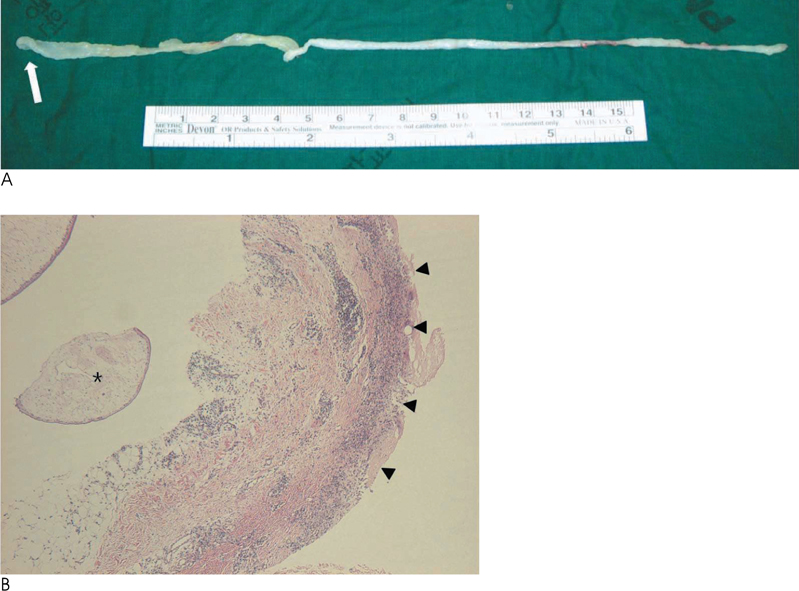
Fig. 4 Photographs of Sparganum obtained by surgical removal. A. An ivory opaque cord-like tapeworm with a scolex (arrow) was extracted in the right breast upper inner quadrant. It measured 24.0×0.7 cm in size. B. Photomicrograph of specimen (original magnification, ×100; hematoxylin-eosin [H-E] stain) show a sparganum larva (asterisk) with the characteristic pattern of noncellular tegument, cellular subtegument, and parenchyma bearing numerous bundles of muscle fibers and excretory canals. A surrounding foreign body granulomatous inflammation and inflammatory cell infiltration such as eosinophils, lymphocytes and histiocytes (arrowheads) are noted. The final pathologic diagnosis was breast sparganosis in the upper inner quadrant of the right breast.
Reference
-
1. Kim HS, Cha ES, Kim HH, Yoo JY. Spectrum of sonographic findings in superficial breast masses. J Ultrasound Med. 2005; 24:663–680.2. Cho JH, Lee KB, Yong TS, Kim BS, Park HB, Ryu KN, et al. Subcutaneous and musculoskeletal sparganosis: imaging characteristics and pathologic correlation. Skeletal Radiol. 2000; 29:402–408.3. Park JH, Chai JW, Cho NR, Paek NS, Guk SM, Shin EH, et al. A surgically confirmed case of breast sparganosis showing characteristic mammography and ultrasonography findings. Korean J Parasitol. 2006; 44:151–156.4. Kim JE, Kim YJ, Kim MY, Han JY. A case of breast sparganosis: MR findings and ultrasonographic findings. Eur J Radiol Extra. 2007; 64:63–65.5. Park HJ, Park NH, Lee EJ, Park CS, Lee SM, Park SI. Ultrasonographic findings of subcutaneous and muscular sparganosis. J Korean Soc Radiol. 2009; 61:183–187.6. Jeong JK, Ryu BY, Lee HW, Kim HK, Choi CS. Sparganosis of the breast. J Korean Surg Soc. 1995; 48:428–432.7. Chung SY, Park KS, Lee Y, Park CK. Breast sparganosis: mammographic and ultrasound features. J Clin Ultrasound. 1995; 23:447–451.8. Moon HG, Jung EJ, Park ST. Breast sparganosis presenting as a breast mass with vague migrating pain. J Am Coll Surg. 2008; 207:292.9. Kim YS, Hwang MS, Lee JK, Kim DS, Lee SK. US findings of breast sparganosis. J Korean Soc Med Ultrasound. 2003; 22:151–156.10. Macura KJ, Ouwerkerk R, Jacobs MA, Bluemke DA. Patterns of enhancement on breast MR images: interpretation and imaging pitfalls. Radiographics. 2006; 26:1719–1734.
- Full Text Links
-
- Actions
-
Cited
- CITED
-
- Close
- Share
-
- Similar articles
-
- Recurrent Ipsilateral Breast Sparganosis: A Case Report
- Sparganosis of the Breast that Mimicked Metastasis: A Case Report
- Sparganosis of the Breast: A Case Report
- Recurrent Breast Sparganosis: Clinical and Radiological Findings
- The Role of Preoperative Breast MRI in Patients With Early-Stage Breast Cancer
