Carcinosarcoma of the Ureter and Urinary Bladder: A Case Report
- Affiliations
-
- 1Department of Radiology, Sanggye Paik Hospital, Inje University College of Medicine, Korea. soohyun@paik.ac.kr
Abstract
- Carcinosarcoma is biphasic neoplasm with distinct carcinomatous and sarcomatous components. Carcinosarcoma arising from the urinary system is extremely rare and only 14 such cases of the ureteral carcinosarcoma have been reported in the medical literature. We experienced a case of surgically proven carcinosarcoma of the ureter and urinary bladder and we report here on the computed tomography findings of this rare neoplasm.
Figure
-
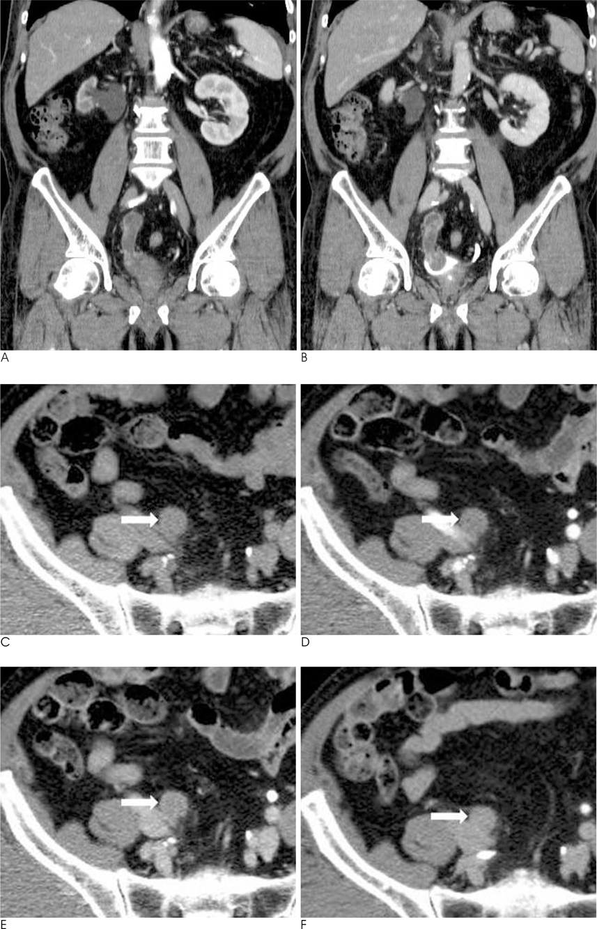
Fig. 1 A 83-year-old man with gross hematuria. A, B. The contrast-enhanced coronal reformatted CT scans show a heterogeneously enhancing intraluminal mass in the right distal ureter and that is protruding into the lumen of the urinary bladder. The attenuation value of the mass was measured as 42 Hounsfield units (HU) during the corticomedullary phase (A) and 66 HU during the nephrographic phase (B). C-F. The axial MDCT scans show different attenuation values of the ureteral mass (arrow) during different phases. The attenuation value of the lesion was measured as 21 HU during the nonenhanced phase (C), 42 HU during the corticomedullary phase (D), 66 HU during the nephrographic phase (E) and 71 HU during the pyelographic phase.
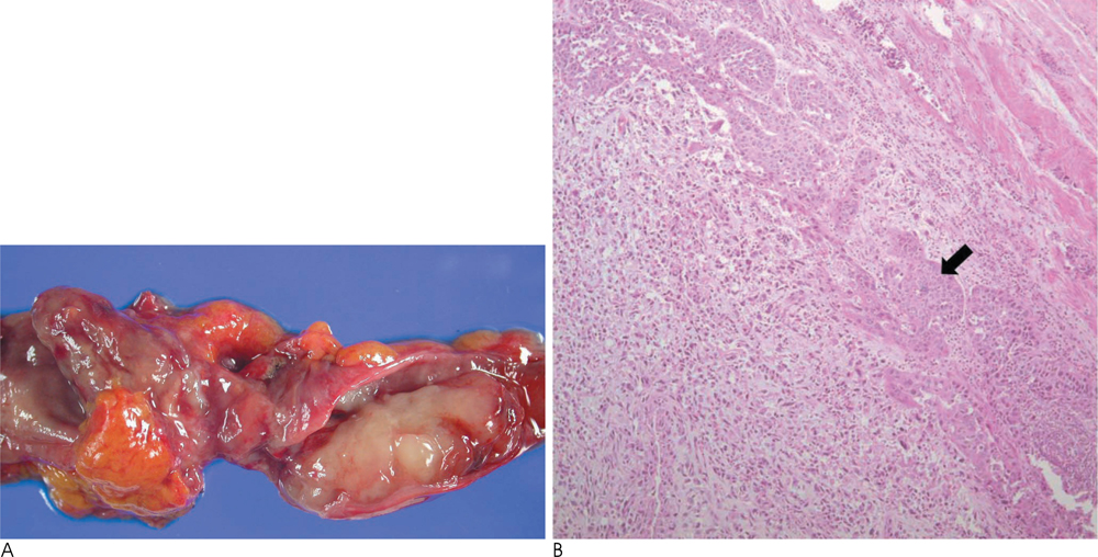
Fig. 2 Pathologic findings of the right ureteral and urinary bladder mass. A. Photograph of the resected ureter shows a grayish yellow mass protruding into the lumen of the distal ureter. B. Photomicrograph of the histological specimen shows a nest of squamous cell carcinoma (arrow) in sarcomatous stroma (H & E, ×10).
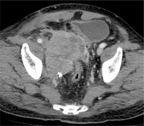
Fig. 3 Two months after the surgery, the CT scan shows a large heterogeneous mass in the right side of the pelvic cavity.
Reference
-
1. Perret L, Chaubert P, Hessler D, Guillou L. Primary heterologous carcinosarcoma (metaplastic carcinoma) of the urinary bladder: a clinicopathologic, immunohistochemical, and ultrastructural analysis of eight cases and a review of the literature. Cancer. 1998; 82:1535–1549.2. Tekes A, Kamel IR, Szarf G, Chan TY, Schoenberg MP, Bluemke DA. Carcinosarcoma of the urinary bladder: dynamic contrast-enhanced MR imaging with clinical and pathologic correlation. AJR Am J Roentgenol. 2003; 181:139–142.3. Perimenis P, Athanasopoulos A, Geragthy J, Speakman M. Carcinosarcoma of the ureter : a rare, pleomorphic, aggressive malignancy. Int Urol Nephrol. 2003; 35:491–493.4. Sumi Y, Shindoh N, Kimizuka T, Katayama H. Carcinosarcoma of the urinary bladder. AJR Am J Roentgenol. 1999; 172:767–769.5. Yilmaz E, Birlik B, Arican Z, Guney S. Carcinosarcoma of the renal pelvis and urinary bladder: a case report. Korean J Radiol. 2003; 4:255–259.6. Baschinsky DY, Chen JH, Vadmal MS, Lucas JG, Bahnson RR, Niemann TH. Carcinosarcoma of the urinary bladder--an aggressive tumor with diverse histogenesis. A clinicopathologic study of 4 cases and review of the literature. Arch Pathol Lab Med. 2000; 124:1172–1178.7. Darko A, DAS K, Bhalla RS, Heller D. Carcinosarcoma of the ureter: report of a case with unusual histology and review of the literature. Int J Urol. 2006; 13:1528–1531.8. Fritz GA, Schoellnast H, Deutschmann HA, Quehenberger F, Tillich M. Multiphasic multidetector-row CT (MDCT) in detection and staging of transitional cell carcinomas of the upper urinary tract. Eur Radiol. 2006; 16:1244–1252.
